. 2025 Aug 17;26(16):7939.
doi: 10.3390/ijms26167939.

Preliminary Preclinical Evaluation of Innovative Bone Scaffolds Composed of Natural Sources-Whey Protein Isolate and Pearl Powder

Affiliations

Preliminary Preclinical Evaluation of Innovative Bone Scaffolds Composed of Natural Sources-Whey Protein Isolate and Pearl Powder

Daniel K Baines et al. Int J Mol Sci. .

Abstract

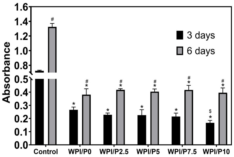
The aim of this work was to produce bone scaffolds containing whey protein isolate and pearl powder and to conduct a preliminary assessment of the biomedical potential in vitro and in vivo. This included analysis of structural, physicochemical, mechanical, and biological properties, which revealed that biomaterials containing pearl powder exhibited an enhanced porous structure, increasing absorptive properties, and decreasing proteolytic capacity with increasing inorganic component content. Pearl powder content in the biomaterials did not clearly influence their mechanical properties or their ability to release calcium ions, as well as proteins. Extracts obtained from all tested biomaterials showed no cytotoxicity in vitro. The surfaces of all biomaterials promoted normal human osteoblast growth, proliferation, and osteogenic differentiation. Furthermore, all biomaterials did not display toxicity in vivo, but no changes in Danio rerio were observed after evaluation of the biomaterial containing the highest amount of pearl powder-10% v/w (marked as WPI/P10). Taking all the obtained results into account, it appears that this biomaterial can be promising for bone scaffolds and similar applications, thanks to its porous structure, high cytocompatibility in vitro, and lack of toxicity in vivo. However, advanced studies will be conducted in the future.

Keywords: Danio rerio; bone; cell-biomaterial interactions; pearl powder; scaffold; whey protein isolate.


Conflict of interest statement

The authors declare no conflicts of interest.

Figures

Figure 1
Figure 1
SEM images presenting microstructure of stiff but flexible hydrogels, Magn. 3000×, scale bar = 1 μm (A) and stereoscopic microscope images presenting macrostructure of stiff but flexible hydrogels (B), Magn. 1.2.×, scale bar = 1 mm and Magn. 3×, scale bar = 500 μm.
Figure 2
Figure 2
FTIR analysis of the WPI/Pearl hydrogels (intensity in arbitrary units (a.u.) with wavenumber). The samples increase in pearl concentration in an ascending order, with the WPI/P0 control, with no pearl powder (black) at the bottom.
Figure 3
Figure 3
Results of swelling assays. The WPI/Pearl and WPI/P0 biomaterial samples were incubated at pH 7 for 5 days. The ratio mass change as a percentage was calculated. Each bar represents the mean ± SD of n = 10. *** p < 0.001–statistically significant results compared to the WPI/P0 base biomaterial (unpaired Student t-test, GraphPad Prism 10, Version 10.4.1 Software).
Figure 4
Figure 4
The results of the mechanical testing for the WPI/Pearl hydrogel. The Young’s modulus (A), the compressive strength (B), and the strain at break (C) were analyzed for each concentration sample group. Each bar represents the mean ± SD of n = 5. *** p < 0.001–statistically significant results compared to the WPI/P0 base biomaterial (unpaired Student t-test, GraphPad Prism 10, Version 10.4.1 Software).
Figure 5
Figure 5
The results of the enzymatic degradation of the WPI/Pearl hydrogel samples. The samples were incubated at pH 7 with proteases for 5 days. The WPI0C sample group is a control without enzymes, whereas the WPI0 is the 40% WPI hydrogel control with enzymes in the solution. Each bar represents the mean ± SD of n = 10 (*** p < 0.001, **** p < 0.0001) compared to the WPI/P0C, one-way ANOVA, followed by Tukey’s multiple comparison test, GraphPad Prism 10, Version 10.4.1 Software).
Figure 6
Figure 6
The ability of biomaterials to absorb or release calcium ions over time. During the experiment, liquid extracts from biomaterials (n = 4) were harvested on day 3, and then a new portion of culture medium was added. The extracts were taken once again on day 6. *-Statistically significant difference compared to the control extract–culture medium incubated without biomaterials and collected at the specified time point; #-statistically significant difference compared to the extract from base biomaterial (WPI/P0), collected at the specified time point; $-statistically significant difference to the extract from WPI/P2.5 biomaterial, collected at the specified time point; &-statistically significant difference compared to the extract from WPI/P7.5 biomaterial, collected at the specified time point. Two-way ANOVA, followed by Bonferroni comparison test, p < 0.05, GraphPad Prism 10, Version 10.4.1 Software.
Figure 7
Figure 7
The ability of biomaterials to release protein over time. During the experiment, liquid extracts from biomaterials (n = 4) were harvested on day 3, and then a new portion of culture medium was added. The extracts were taken once again on day 6. *-Statistically significant difference compared to the control extract–culture medium incubated without biomaterials and collected at the specified time point; #-statistically significant difference compared to the extract from base biomaterial (WPI/P0), collected at the specified time point; $-statistically significant difference to the extract from WPI/P2.5 biomaterial, collected at the specified time point; ^-statistically significant difference compared to the extract from WPI/P5 biomaterial, collected at the specified time point. Two-way ANOVA, followed by Bonferroni comparison test, p < 0.05, GraphPad Prism 10, Version 10.4.1 Software).
Figure 8
Figure 8
Viability of normal human osteoblasts treated with biomaterial extracts (n = 4) for 24 h. Cell viability was determined by the MTT assay. No statistically significant differences were observed between groups (p > 0.05, based on one-way ANOVA, followed by Tukey’s multiple comparison test, p < 0.05, GraphPad Prism 10, Version 10.4.1 Software).
Figure 9
Figure 9
Viability of normal human osteoblasts growing on control biomaterials–polystyrene and tested biomaterials composed of WPI without or with pearl powder (WPI/P0 and WPI/P2.5; WPI/P5; WPI/P7.5; WPI/P10, respectively). After 48 h of incubation, cells were differentially stained using the Live/Dead Double Cell Staining Kit and observed using a confocal laser scanning microscope (CLSM). Only living cells were observed, which emitted green fluorescence (if dead cells were present, they would emit red fluorescence). Magnification = 100×, bar scale = 150 μm.
Figure 10
Figure 10
Quantitative assessment of proliferation of normal human osteoblasts cultured on control biomaterials–polystyrene (n = 4) and tested biomaterials (n = 4) composed of WPI without or with pearl powder (WPI/P0 and WPI/P2.5; WPI/P5; WPI/P7.5; WPI/P10, respectively). After 3 and 6 days of incubation, cell metabolic activity was assessed using the WST-8 assay. The OD value is directly proportional to the number of viable, metabolically active cells. *–Statistically significant difference compared to the control biomaterial–polystyrene at the specified time point; #–statistically significant difference compared to the results obtained after 3 and 6 days of incubation; $-statistically significant difference between WPI/P0 and WPI/P10 biomaterial on day 3. Two-way ANOVA, followed by Bonferroni comparison test, p < 0.05, GraphPad Prism 10, Version 10.4.1 Software).
Figure 11
Figure 11
Qualitative assessment of proliferation of normal human osteoblasts cultured on control biomaterials–polystyrene and tested biomaterials composed of WPI without or with pearl powder (WPI/P0 and WPI/P2.5; WPI/P5; WPI/P7.5; WPI/P10, respectively). After 3 and 6 days of incubation, cells were stained using the Hoechst 33342 and AlexaFluorTM 635 Phalloidin and observed using a confocal laser scanning microscope (CLSM). Live cells = green fluorescence; dead cells = red fluorescence. Magnification = 200×, bar scale = 150 μm.
Figure 12
Figure 12
Qualitative assessment of osteogenic differentiation of normal human osteoblasts cultured on control biomaterials–polystyrene and tested biomaterials composed of WPI without or with pearl powder (WPI/P0 and WPI/P2.5; WPI/P5; WPI/P7.5; WPI/P10, respectively). After 21 days of incubation, cells were incubated with primary anti-collagen I antibody or primary anti-osteocalcin antibody, followed by staining with specified secondary antibody-conjugated with Alexa Fluor 488 and additionally with Hoechst 33342. Then, the cells were observed using a confocal laser scanning microscope (CLSM). Cell nuclei = blue fluorescence; collagen or osteocalcin = green fluorescence. Magnification = 200× or 400×, bar scale = 70 or 30 μm.
Figure 13
Figure 13
Heart rate at 96 hpf in the FET test. Heart rate was measured manually for exactly 10 s and multiplied by 6, results presented as mean with SD, n = 12. The results were not statistically significant among themselves, based on one-way ANOVA, followed by Tukey’s multiple comparison test, p < 0.05, GraphPad Prism 10, Version 10.4.1 Software).
Figure 14
Figure 14
Distribution of Malformed vs. Non-Malformed larvae. Malformation was defined as pericardial oedema.
Figure 15
Figure 15
Zebrafish larvae in lateral position at 96 hpf of the FET test. A red asterisk (*) indicates the presence of pericardial oedema, also shown in the right panel with a red arrow. Images were taken using a SteREO Discovery.V8 microscope (Carl Zeiss Microscopy GmbH, Hallbergmoos, Germany) at 3.2× magnification.
Figure 16
Figure 16
Heatmap of Bonferroni-corrected p-values from Fisher’s exact test (2 × 2) for all group pairs.
Figure 17
Figure 17
An image of the WPI/Pearl hydrogels post-sterilization. From left to right–WPI/P0, WPI/P2.5, WPI/P5, WPI/P7.5 and WPI/P10.

Similar articles

References

    1. Zhou Z., Feng W., Moghadas B.K., Baneshi N., Noshadi B., Baghaei S., Dehkordi D.A. Review of recent advances in bone scaffold fabrication methods for tissue engineering for treating bone diseases and sport injuries. Tissue Cell. 2024;88:102390. doi: 10.1016/j.tice.2024.102390. - DOI - PubMed
    1. Lee S.S., Du X., Kim I., Ferguson S.J. Scaffolds for bone-tissue engineering. Matter. 2022;5:2722–2759. doi: 10.1016/j.matt.2022.06.003. - DOI
    1. Wu A.M., Bisignano C., James S.L., Abady G.G., Abedi A., Abu-Gharbieh E., Alhassan R.K., Alipour V., Arabloo J., Asaad M., et al. Global, regional, and national burden of bone fractures in 204 countries and territories, 1990–2019: A systematic analysis from the Global Burden of Disease Study 2019. Lancet Heal. Longev. 2021;2:e580–e592. doi: 10.1016/S2666-7568(21)00172-0. - DOI - PMC - PubMed
    1. Yazdanpanah Z., Johnston J.D., Cooper D.M.L., Chen X. 3D Bioprinted Scaffolds for Bone Tissue Engineering: State-of-the-Art and Emerging Technologies. Front. Bioeng. Biotechnol. 2022;10:824156. doi: 10.3389/fbioe.2022.824156. - DOI - PMC - PubMed
    1. Percival K.M., Paul V., Husseini G.A. Recent Advancements in Bone Tissue Engineering: Integrating Smart Scaffold Technologies and Bio-Responsive Systems for Enhanced Regeneration. Int. J. Mol. Sci. 2024;25:6012. doi: 10.3390/ijms25116012. - DOI - PMC - PubMed

Grants and funding

LinkOut - more resources