. 2025 Aug 19;26(16):8012.
doi: 10.3390/ijms26168012.

25-Hydroxycholesterol Induces Intrinsic Apoptosis via Mitochondrial Pathway in BE(2)-C Human Neuroblastoma Cells

Affiliations

25-Hydroxycholesterol Induces Intrinsic Apoptosis via Mitochondrial Pathway in BE(2)-C Human Neuroblastoma Cells

Jaesung Kim et al. Int J Mol Sci. .

Abstract

An oxysterol, 25-Hydroxycholesterol (25OHChol), is produced through cholesterol oxidation and is involved in various cellular processes, including apoptosis. However, the precise mechanisms underlying 25OHChol-induced apoptosis in neuroblastoma cells remain unclear. The aim of this study was to elucidate the detailed molecular mechanisms by which 25OHChol induces apoptosis in human neuroblastoma cells. This study explores the apoptotic effects of 25OHChol and the associated signaling pathways in BE(2)-C cells, a widely used human neuroblastoma cell model for neuronal differentiation and cancer research. To evaluate the cytotoxicity of 25OHChol, cell viability was assessed using the CCK-8 assay, which demonstrated a concentration-dependent decline, indicating a potential induction of cell death. Morphological changes characteristic of apoptosis, such as nuclear condensation and fragmentation, were confirmed via DAPI staining. Additionally, Annexin V/PI flow cytometry analysis revealed an increase in late apoptotic cell populations, further corroborating apoptosis induction. To investigate the molecular mechanisms, we analyzed the expression of Bcl-2 family proteins via Western blotting. The results showed an elevated Bax/Bcl-2 ratio, suggesting activation of the intrinsic mitochondrial apoptotic pathway. This was further supported by a reduction in mitochondrial membrane potential (MMP), as measured by flow cytometry. Increased caspase-9 and caspase-3/7 activity provided additional evidence for caspase-mediated apoptosis. Moreover, treatment with the pan-caspase inhibitor Z-VAD-FMK led to a dose-dependent increase in cell viability, confirming the essential role of caspases in 25OHChol-induced apoptosis. In conclusion, this study demonstrates that 25OHChol triggers apoptosis in BE(2)-C neuroblastoma cells through activation of the intrinsic mitochondrial apoptotic pathway. These findings provide new insights into the cytotoxic effects of 25OHChol and its potential role in neuroblastoma cell death.

Keywords: 25-hydroxycholesterol; mitochondria; neuro-inflammation; neuronal cell death; oxysterol.


Conflict of interest statement

The authors declare no conflicts of interest.

Figures

Figure 1
Figure 1
Effects of 25OHChol on viability of BE(2)-C cells. (A) BE(2)-C cells were treated with 1 μg/mL of Chol or oxysterols for 48 h. The cells were incubated at indicated concentrations of 25OHChol for 48 h, and at 1 μg/mL of the oxysterol for indicated times (B). The cell viability was measured using the CCK-8 assay, and data are presented as the mean ± SD, with three replicates per condition (n = 3). * p < 0.05, ** p < 0.01, and *** p < 0.001 compared to the control group.
Figure 2
Figure 2
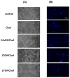
Effects of 25OHChol on morphological changes in BE(2)-C cells. BE(2)-C cells were serum-starved and then incubated for 48 h with Chol or oxysterols (1 µg/mL). (A) Morphological changes in BE(2)-C cells were assessed using a light microscope at 200× magnification. (B) Nuclear morphological changes were evaluated by staining the cells with DAPI and observing them under a confocal microscope at 400× magnification.
Figure 3
Figure 3
Effects of 25OHChol on apoptosis of BE(2)-C cells. BE(2)-C cells were serum-starved and then incubated for 48 h with Chol or oxysterols (1 µg/mL). Unstained and single-stained samples with Annexin V and PI were prepared to establish compensation. The cells were then double-stained with Chol or oxysterols and analyzed by flow cytometry.
Figure 4
Figure 4
Expression of Bax and Bcl-2 proteins after treatment with 25OHChol in BE(2)-C cells. BE(2)-C cells were serum-starved and then incubated with 25OHChol (1 µg/mL) for 0, 24, and 48 h. (A) Cells were lysed, and total protein was analyzed via Western blot to detect Bax and Bcl-2. The band intensity ratio of Bax and Bcl-2 was calculated and presented using ImageJ software (ver.1.54p; National Institutes of Health, Bethesda, MD, USA). (B) The mean values of Bax/Bcl-2 ratio displayed as a graph and data are presented as the mean ± SD, with three replicates per condition (n = 3). * p < 0.05 compared to the control group.
Figure 5
Figure 5
Effects of mitochondrial membrane potential (MMP) after treatment with 25OHChol in BE(2)-C cells. BE(2)-C cells were treated with Chol or oxysterols at a concentration of 1 µg/mL for 48 h. Following treatment, the cells were trypsinized and stained with JC-1 dye at 37 °C for 20 min. Mitochondrial membrane potential was then assessed using flow cytometry. (A) shows data from individual independent experiments, while (B) represents the mean values of cells with a loss of mitochondrial membrane potential. Data are presented as the mean ± SD from three replicates per condition (n = 3). ** p < 0.01 compared to the control group.
Figure 6
Figure 6
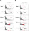
Effects of 25OHChol on caspase activity in BE(2)-C cells. BE(2)-C cells were treated with Chol or oxysterols at a concentration of 1 µg/mL for 48 h, after which caspase activity was evaluated using flow cytometry and Western blotting. (A) To measure caspase-9 activity, cells were incubated with FITC-LEHD-FMK at 37 °C with 5% CO2 for 1 h, followed by flow cytometry analysis. (B) For assessing caspase-3/7 activity, cells were incubated with TF2-DEVD-FMK under the same conditions for 3 h, and activity was then measured by flow cytometry. (C) In a separate experiment, BE(2)-C cells were treated with 25OHChol at 1 µg/mL, and cell lysates were harvested at 0, 24, and 48 h after treatment. Total protein was analyzed via Western blot to detect caspase-3. The band intensity ratio of cleaved caspase-3/pro-caspase-3 was calculated and the mean values presented as a graph, with data shown as the mean ± SD from three replicates per condition (n = 3). Statistical significance was determined at *** p < 0.001 compared to the control group. (D) BE(2)-C cells were exposed to 1 μg/mL 25OHChol and Z-VAD-FMK at specified concentrations for 48 h. Cell viability was determined using the CCK-8 assay, with data presented as the mean ± SD from three replicates per condition (n = 3). *** p < 0.001 compared to the control group and # p < 0.5, ## p < 0.1, ### p < 0.01 compared to the 25OHChol group.
Figure 6
Figure 6
Effects of 25OHChol on caspase activity in BE(2)-C cells. BE(2)-C cells were treated with Chol or oxysterols at a concentration of 1 µg/mL for 48 h, after which caspase activity was evaluated using flow cytometry and Western blotting. (A) To measure caspase-9 activity, cells were incubated with FITC-LEHD-FMK at 37 °C with 5% CO2 for 1 h, followed by flow cytometry analysis. (B) For assessing caspase-3/7 activity, cells were incubated with TF2-DEVD-FMK under the same conditions for 3 h, and activity was then measured by flow cytometry. (C) In a separate experiment, BE(2)-C cells were treated with 25OHChol at 1 µg/mL, and cell lysates were harvested at 0, 24, and 48 h after treatment. Total protein was analyzed via Western blot to detect caspase-3. The band intensity ratio of cleaved caspase-3/pro-caspase-3 was calculated and the mean values presented as a graph, with data shown as the mean ± SD from three replicates per condition (n = 3). Statistical significance was determined at *** p < 0.001 compared to the control group. (D) BE(2)-C cells were exposed to 1 μg/mL 25OHChol and Z-VAD-FMK at specified concentrations for 48 h. Cell viability was determined using the CCK-8 assay, with data presented as the mean ± SD from three replicates per condition (n = 3). *** p < 0.001 compared to the control group and # p < 0.5, ## p < 0.1, ### p < 0.01 compared to the 25OHChol group.

References

    1. Schulte J., Schulte S., Heukamp L., Astrahantseff K., Stephan H., Fischer M., Schramm A., Eggert A. Targeted therapy for neuroblastoma: ALK inhibitors. Klin. Pädiatrie. 2013;225:303–308. doi: 10.1055/s-0033-1357132. - DOI - PubMed
    1. Esposito M.R., Aveic S., Seydel A., Tonini G.P. Neuroblastoma treatment in the post-genomic era. J. Biomed. Sci. 2017;24:14. doi: 10.1186/s12929-017-0319-y. - DOI - PMC - PubMed
    1. Ahmed S., Goel S., Khandwala M., Agrawal A., Chang B., Simmons I. Neuroblastoma with orbital metastasis: Ophthalmic presentation and role of ophthalmologists. Eye. 2006;20:466–470. doi: 10.1038/sj.eye.6701912. - DOI - PubMed
    1. Costa A.D., Zerbini M.C.N., Cristofani L. Metastatic congenital neuroblastoma associated with in situ neuroblastoma: Case report and review of literature. Autops. Case Rep. 2014;4:27. doi: 10.4322/acr.2014.017. - DOI - PMC - PubMed
    1. Maris J.M. Recent advances in neuroblastoma. N. Engl. J. Med. 2010;362:2202–2211. doi: 10.1056/NEJMra0804577. - DOI - PMC - PubMed

MeSH terms

Substances