

Lateral Geniculate Nucleus Volume Assessed by 7 Tesla MRI 3D MT-Weighted SILENT Protocol in Patients with STARGARDT Disease-Pilot Study
- PMID: 40869492
- PMCID: PMC12386881
- DOI: 10.3390/jcm14165666
Lateral Geniculate Nucleus Volume Assessed by 7 Tesla MRI 3D MT-Weighted SILENT Protocol in Patients with STARGARDT Disease-Pilot Study
Abstract
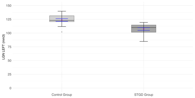
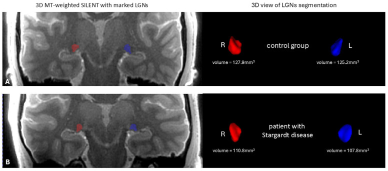
Background/Objectives: To quantitatively assess lateral geniculate nucleus (LGN) volume using 7 Tesla MRI in patients with Stargardt disease (STGD). Methods: A total of 18 patients with STGD and 15 healthy volunteers were examined with a 7 Tesla MRI of the brain. Measures of LGN volume were performed manually by three independent investigators (radiologists) using ITK-SNAP software, version 4.0.0-rc.2. The volume of the thalamus was evaluated using the open-source automated software package FreeSurfer. Before 7 Tesla MRI, patients underwent ophthalmic examination and 1.5 Tesla MRI. Results: The average LGN volume in both hemispheres was significantly smaller in patients with STGD (right, -111.2 mm3; left, 107.4 mm3) than in the control group (right, -128.7 mm3; left, 123.6 mm3, respectively) (p < 0.0001). The ratio of LGN to thalamus in the right hemisphere was significantly lower (p = 0.024) in the group of patients with STGD (0.014) than in the control group (0.017). Conclusions: The right and left LGN volumes in MR 7T imaging, as well as the right LGN/thalamus ratio, were reduced in patients with STGD compared to controls. 7T MRI using the 3D MT-weighted SILENT protocol provides new insight into structural changes in the brain in retinal dystrophies and offers a possible marker of the response to future therapies in STGD.
Keywords: 7 Tesla MRI; Stargardt disease; lateral geniculate nucleus.
Conflict of interest statement
The authors declare no conflicts of interest.
Figures


References
-
- Huang X., Ruan G., Zhang J., Luo F., Zhao K. Genetic Diagnosis of Familial Exudative Vitreoretinopathy by Targeted Next-Generation Sequencing. Ophthalmic Genet. 2021;42:196–202.
-
- Radu R.A., Han Y., Bui T.V., Nusinowitz S., Bok D., Lichter J.B., Travis G.H., Mata N.L. Reductions in Serum Vitamin A in Stargardt’s Patients Treated with Oral Fenretinide. Investig. Ophthalmol. Vis. Sci. 2011;53:4409–4415.
-
- Tsang H.S., Sharma T. Stargardt Disease. Adv. Exp. Med. Biol. 2018;1085:139–151. - PubMed
Grants and funding
LinkOut - more resources
Full Text Sources
