Review
. 2025 Aug 18;14(16):5821.
doi: 10.3390/jcm14165821.

Aggressiveness in Well-Differentiated Small Intestinal Neuroendocrine Tumors: A Rare Case and Narrative Literature Review

Affiliations
Review

Aggressiveness in Well-Differentiated Small Intestinal Neuroendocrine Tumors: A Rare Case and Narrative Literature Review

Laurențiu Augustus Barbu et al. J Clin Med. .

Abstract

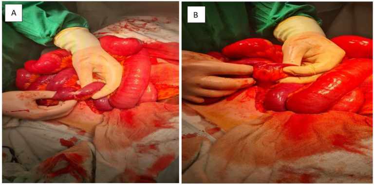
Background: Small intestinal neuroendocrine tumors (SI-NETs) are the most common malignancies of the small bowel. Although typically well differentiated and slow-growing, they may exhibit aggressive behavior, especially when diagnosed at an advanced stage. Objective: To illustrate the diagnostic and therapeutic challenges of advanced SI-NETs through a rare case presentation and a narrative review of recent studies in the literature. Methods: A narrative literature review was conducted using the PubMed database to examine the incidence, risk factors, diagnostic modalities, and treatment strategies for advanced-stage SI-NETs. The search included studies published between January 2010 and June 2025 and focused on human subjects, using keywords such as "small intestinal neuroendocrine tumor", "metastasis", "tumor grade", and "treatment". Results: We report the case of a 68-year-old man who presented with bowel obstruction. Imaging and surgical exploration revealed a jejunoileal SI-NET with extensive liver and peritoneal metastases, mesenteric fibrosis, and ascites. Histopathology confirmed a well-differentiated grade 2 tumor (Ki-67: 3%) positive for chromogranin A and CD56. Despite a low proliferative index, the tumor demonstrated aggressive clinical behavior. The patient underwent emergency enterectomy with ileostomy and was referred for further evaluation, including somatostatin receptor imaging and consideration for peptide receptor radionuclide therapy (PRRT). Conclusions: This case highlights the potential for aggressive progression in well-differentiated SI-NETs with low Ki-67 indices. Histological grade alone may not predict clinical behavior. Early diagnosis, comprehensive staging, and individualized multidisciplinary management-guided by functional imaging and receptor profiling-are critical to improving outcomes in advanced SI-NETs.

Keywords: PRRT; SI-NET; carcinoid; chromogranin A; immunohistochemistry; liver metastasis; peritoneal metastasis; small intestinal neuroendocrine tumor.


Conflict of interest statement

The authors declare that there are no conflicts of interest.

Figures

Figure 1
Figure 1
(A,B): Contrast-enhanced abdominal CT. (A) A pelvic mesenteric tumor mass (yellow arrow) is seen causing significant dilatation of adjacent small bowel loops, suggestive of partial obstruction. (B) Multiple hypodense hepatic lesions (yellow arrow) are visualized, consistent with secondary metastatic deposits from a neuroendocrine tumor.
Figure 2
Figure 2
(A,B): Contrast-enhanced abdominal CT. Multiple soft-tissue density nodules (yellow arrow) are diffusely distributed throughout the peritoneal cavity, consistent with metastatic involvement of the parietal peritoneum. The pattern and morphology of these lesions support a diagnosis of peritoneal carcinomatosis (yellow arrow) secondary to a neuroendocrine tumor.
Figure 3
Figure 3
(A,B): Intraoperative view. A markedly stenotic segment of small intestine is shown, with significant proximal bowel dilatation. These findings are consistent with partial obstruction secondary to tumor infiltration and associated mesenteric fibrosis.
Figure 4
Figure 4
Hematoxylin and eosin (H&E) staining (×100). Nests of uniform neuroendocrine tumor cells exhibiting round nuclei, “salt and pepper” chromatin, and minimal cytological atypia, consistent with a well-differentiated morphology.
Figure 5
Figure 5
Chromogranin A immunostaining (×100). Diffuse and strong cytoplasmic staining is observed in nests of uniform neuroendocrine tumor cells, confirming neuroendocrine differentiation.
Figure 6
Figure 6
Chromogranin A immunostaining (×100). Strong and diffuse cytoplasmic positivity is observed in nests of uniform neuroendocrine tumor cells, consistent with neuroendocrine differentiation.
Figure 7
Figure 7
CD56 immunostaining (×100). The image shows nests of well-differentiated neuroendocrine tumor cells exhibiting diffuse and strong membranous CD56 positivity, consistent with neuroendocrine phenotype.
Figure 8
Figure 8
Synaptophysin immunostaining, ×100. Focal and weak immunoreactivity is observed in clusters of monotonous neuroendocrine cells, consistent with variable synaptophysin expression in well-differentiated neuroendocrine tumors.

Similar articles

References

    1. Sun Y., Lohse C., Smyrk T., Hobday T., Kroneman T., Zhang L. The Influence of Tumor Stage on the Prognostic Value of Ki-67 Index and Mitotic Count in Small Intestinal Neuroendocrine Tumors. Am. J. Surg. Pathol. 2018;42:247–255. doi: 10.1097/PAS.0000000000000968. - DOI - PubMed
    1. Kim M.K., Warner R.R., Roayaie S., Harpaz N., Ward S.C., Itzkowitz S., Wisnivesky J.P. Revised staging classification improves outcome prediction for small intestinal neuroendocrine tumors. J. Clin. Oncol. 2013;31:3776–3781. doi: 10.1200/JCO.2013.51.1477. - DOI - PMC - PubMed
    1. Wu L., Fu J., Wan L., Pan J., Lai S., Zhong J., Chung D.C., Wang L. Survival outcomes and surgical intervention of small intestinal neuroendocrine tumors: A population based retrospective study. Oncotarget. 2017;8:4935–4947. doi: 10.18632/oncotarget.13632. - DOI - PMC - PubMed
    1. Mantzoros I., Savvala N.A., Ioannidis O., Parpoudi S., Loutzidou L., Kyriakidou D., Cheva A., Intzos V., Tsalis K. Midgut neuroendocrine tumor presenting with acute intestinal ischemia. World J. Gastroenterol. 2017;23:8090–8096. doi: 10.3748/wjg.v23.i45.8090. - DOI - PMC - PubMed
    1. Modlin I.M., Kidd M., Latich I., Zikusoka M.N., Shapiro M.D. Current status of gastrointestinal carcinoids. Gastroenterology. 2005;128:1717–1751. doi: 10.1053/j.gastro.2005.03.038. - DOI - PubMed

LinkOut - more resources