Review
. 2025 Aug 21;14(16):5895.
doi: 10.3390/jcm14165895.

Uncommon and Accessory Electrocardiographic Findings in Brugada Syndrome: A Review

Affiliations
Review

Uncommon and Accessory Electrocardiographic Findings in Brugada Syndrome: A Review

Antonino Micari et al. J Clin Med. .

Abstract

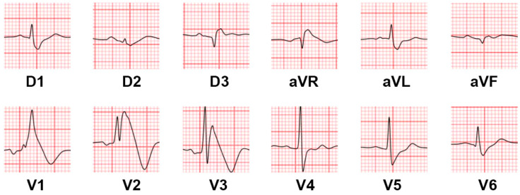
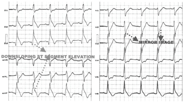
Brugada syndrome (BrS) is a cardiac arrhythmic disorder associated with distinctive electrocardiographic (ECG) abnormalities and an increased risk of sudden cardiac death due to ventricular arrhythmias. While the classic BrS ECG pattern is a coved ST-segment elevation in the right precordial leads, a wide spectrum of atypical ECG presentations can mislead the diagnosis. This review discusses rare and under-recognized ECG findings associated with BrS, including its coexistence with right and left bundle branch block, alterations in peripheral leads and in the morphology of the QRS complex, as well as atrioventricular conduction abnormalities. Emphasis is placed on the clinical relevance of these findings, their underlying electrophysiological mechanisms, and their prognostic implications. Recognizing these atypical manifestations is critical to avoid misdiagnosing or failing to recognize the condition in patients with BrS.

Keywords: Brugada syndrome; electrocardiography; sudden cardiac death; ventricular arrhythmias.


Conflict of interest statement

The authors declare no conflicts of interest.

Figures

Figure 2
Figure 2
(A). Surface ECG showing premature atrial complex conducted with aberration and with a LBBB morphology, masking the Brugada pattern in the precordial leads. (B). Right atrial pacing conducted with aberration showing a wide QRS with LBBB morphology, which hides the Brugada pattern normally visible in the precordial leads with narrow QRS. Adapted with permission from Arana-Rueda E. et al. [20].
Figure 1
Figure 1
Basal 12-lead surface ECG showing a RBBB with a wide terminal R’ wave in right precordial leads, suggestive of a Brugada type 1 pattern.
Figure 3
Figure 3
Basal 12-lead surface ECG showing a ST-depression in inferior leads, with an apparently normal ST segment in the right precordial leads at the fourth intercostal space. The precordial leads at the second intercostal space show a type 1 Brugada pattern with a wide terminal R’ wave. The basal ECG allowed us to suspect a Brugada pattern only through the analysis of the inferior leads.
Figure 4
Figure 4
A 12-lead ECG showing a downsloping ST-segment elevation in inferior leads with a prominent J wave. A mirror image can be seen in anterior precordial leads (V2–V3). Reproduced with permission from Riera et al. [30].
Figure 5
Figure 5
(A). ECG recorded in a 61-year-old male who experienced syncope at night. VF was detected 57 months after ICD implantation. hV1–hV6 are placed one intercostal space higher than the regular lead positions (V1–V6). Multiple R waves existed in leads II, III, aVF, V1, hV1, and hV2, and fragmented QRS were present in leads reflecting alteration in the inferior and RVOT regions. (BD). Progressive increments of QRS fragmentation (arrowheads indicate new QRS spikes that appeared during follow-up). (E). Effect of epicardial ablation of the right ventricle: Before ablation, five spikes (arrowheads) were observed within QRS complex in lead V2. After ablation, only three spikes (arrowheads) can be seen, with a reduction in ST-segment elevation. Reproduced with permission from Morita et al. [48].
Figure 6
Figure 6
ECG from a 40-year-old patient with pathogenic mutation affecting the SCN5A gene, showing a first-degree atrioventricular block associated with a Brugada pattern. In the inferior leads, a slight notching of the P wave is also noted, which is prolonged, indicative of a possible intra-atrial conduction delay.

References

    1. Hiss R.G., Lamb L.E. Electrocardiographic findings in 122,043 individuals. Circulation. 1962;25:947–961. doi: 10.1161/01.CIR.25.6.947. - DOI - PubMed
    1. Fleg J.L., Das D.N., Lakatta E.G. Right bundle branch block: Long-term prognosis in apparently healthy men. J. Am. Coll. Cardiol. 1983;1:887–892. doi: 10.1016/S0735-1097(83)80204-6. - DOI - PubMed
    1. Aizawa Y., Takatsuki S., Kimura T., Nishiyama N., Fukumoto K., Tanimoto Y., Tanimoto K., Miyoshi S., Suzuki M., Yokoyama Y., et al. Ventricular fibrillation associated with complete right bundle branch block. Heart Rhythm. 2013;10:1028–1035. doi: 10.1016/j.hrthm.2013.03.013. - DOI - PMC - PubMed
    1. Bussink B.E., Holst A.G., Jespersen L., Deckers J.W., Jensen G.B., Prescott E. Right bundle branch block: Prevalence, risk factors, and outcome in the general population: Results from the Copenhagen City Heart Study. Eur. Heart J. 2013;34:138–146. doi: 10.1093/eurheartj/ehs291. - DOI - PubMed
    1. Gaba P., Pedrotty D., DeSimone C.V., Bonikowske A.R., Allison T.G., Kapa S. Mortality in Patients With Right Bundle-Branch Block in the Absence of Cardiovascular Disease. J. Am. Heart Assoc. 2020;9:e017430. doi: 10.1161/JAHA.120.017430. - DOI - PMC - PubMed

LinkOut - more resources