. 2025 Jul 24;16(8):861.
doi: 10.3390/genes16080861.

Construction of a Genetic Prognostic Model in the Glioblastoma Tumor Microenvironment

Affiliations

Construction of a Genetic Prognostic Model in the Glioblastoma Tumor Microenvironment

Wenhui Wu et al. Genes (Basel). .

Abstract

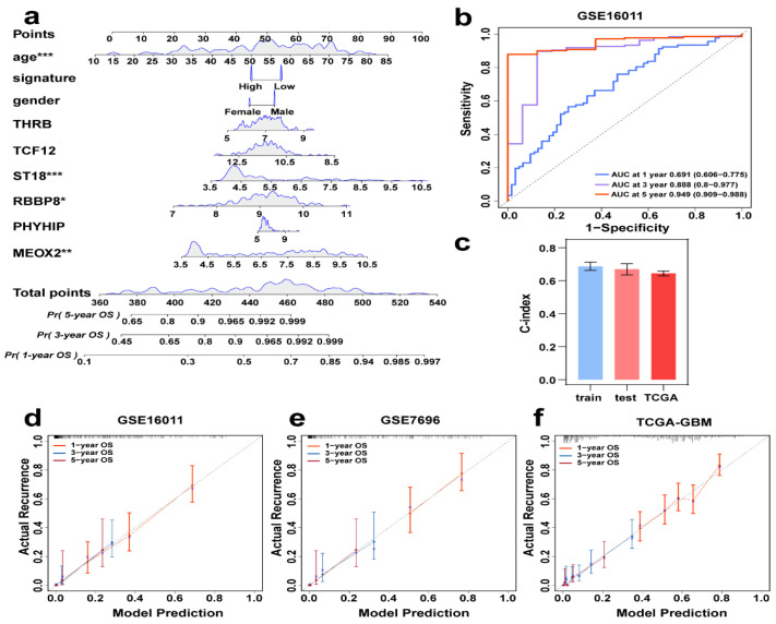
Background: Glioblastoma (GBM) is one of the most challenging malignancies in all of neoplasms. These malignancies are associated with unfavorable clinical outcomes and significantly compromised patient wellbeing. The immunological landscape within the tumor microenvironment (TME) plays a critical role in determining GBM prognosis. By mining data from The Cancer Genome Atlas (TCGA) and Gene Expression Omnibus (GEO) databases and correlating them with immune responses in the TME, genes associated with the immune microenvironment with potential prognostic value were obtained. Method: We selected GSE16011 as the training set. Gene expression profiles were substrates scored by both ESTIMATE and xCell, and immune cell subpopulations in GBM were analyzed by CIBERSORT. Gene expression profiles associated with low immune scores were performed by lasso regression, Cox analysis and random forest (RF) to identify a prognostic model for the multiple genes associated with immune infiltration in GBM. Then we constructed a nomogram to optimize the prognostic model using GSE7696 and TCGA-GBM as validation sets and evaluated these data for gene mutation and gene enrichment analysis.

Result: The prognostic correlation between the six genes (MEOX2, PHYHIP, RBBP8, ST18, TCF12, and THRB) and GBM was finally found by lasso regression, Cox regression, and RF, and the online database obtained that all six genes were differentially expressed in GBM. Therefore, a prognostic correlation model was constructed based on the six genes. Kaplan-Meier (KM) survival analysis showed that this prognostic model had excellent prognostic ability.

Conclusions: Prognostic models based on tumor microenvironment and immune score stratification and the construction of related genes have potential applications for prognostic analysis of GBM patients.

Keywords: glioblastoma; nomogram; prognosis; tumor microenvironment.


Conflict of interest statement

The authors declare no conflicts of interest.

Figures

Figure 1
Figure 1
Immune landscape characterization in GBM. (a) CIBERSORT-derived histogram of 22 immune cell fractions in GBM. (b) Immune cell interaction network in GBM. (c) Heatmap depicting immune cell distribution across GBM cases. (d) Cumulative plots of ESTIMATE and xCell-calculated immune/stromal scores in the GBM cohort.
Figure 2
Figure 2
GSE16011 GBM survival analysis via Kaplan–Meier by score type. (a) Immune scores; (b) stromal; (c) estimates scores; (d) tumor purity scores.
Figure 3
Figure 3
Construction of risk model based on GBM cohort: (a) Heatmap showing gene distribution and clinical information of the cohort; (b) Volcano plot showing differential gene expression. Up (red): upregulated genes (logFC > 2, p-value < 0.05); Down (blue): downregulated genes (logFC < −2, p-value < 0.05); Not (grey): non-significant genes. (c,d) Llasso Cox model analysis identified 22 genes. The different colors of the curves represent the changes in coefficients of different genes. (e) RF analysis (left) evaluated prediction error rates across decision trees to determine model convergence , while variable importance scores (right) identified the top 14 survival-associated genes (final selected MEOX2, PHYHIP, RBBP8, ST18, TCF12, and THRB, highlighted in red).
Figure 4
Figure 4
Risk score curves, survival time and survival status scatter plots, and clustering heat maps for (a) GSE16011, (b) GSE7696, and (c) TCGA-GBM.
Figure 5
Figure 5
Risk score-based KM survival curves for the patient cohorts (a) GSE16011, (b) GSE7696, and (c) TCGA-GBM. (d) Protein expression of MEOX2, PHYHIP, RBBP8, ST18, TCF12, and THRB in tumor tissues in the HPA database.
Figure 6
Figure 6
Individual integrated prognostic model construction. (a) A prognostic nomogram consisting of clinical information and genes together, predicting survival by score for different years. * p < 0.05, ** p < 0.01, *** p < 0.001. (b) GSE16011Time-ROC curves of 1, 3, and 5 years AUC. (c) C-index values of training (GSE16011), test (GSE7696), and TCGA data. (df) Calibration curves of GSE16011, GSE7696, and TCGA-GBM, where the red, blue, and purple lines are the one-year, three-year, and five-year projections, respectively.
Figure 7
Figure 7
Top 10 significantly enriched functional terms. (a) biological process (BP) (b) cellular component (CC) (c) molecular functions (MFs) of GO analysis. (d) KEGG-Gene-Concept Network of top five categories.
Figure 8
Figure 8
Mutational profiles across TCGA-GBM cases. (a) Oncoplot displaying the top 20 most recurrently mutated genes per variant class. (b) The gene cloud shows the total number of samples of each mutated gene as a proportion of the total number of mutations, and the size of each gene is proportional to the total number of samples in which it is mutated. (c) The correlation graph shows the mutually exclusive or coexisting status of the most mutated top genes. The green color is for mutated genes that tend to coexist; the yellow color is for mutually exclusive genes, and the shade of color indicates the significance. (d) Variant allele frequency (VAF) distribution in mutants (box plot). (e) Top 10 significantly enriched oncogenic pathways with sample prevalence rates. (f) Prognostic impact of mutant gene sets (KM analysis).
Figure 9
Figure 9
Correlation analysis of gene tagging with tumor purity and immune cell infiltration in GBM cancer: (a) MEOX2, (b) PHYHIP, (c) RBBP8, (d) ST18, (e) TCF12, and (f) THRB. Each black dot represents a case.
Figure 10
Figure 10
Differential expression of genes between normal and cancerous tissues (a) MEOX2, (b) PHYHIP, (c) RBBP8, (d) ST18, (e) TCF12, and (f) THRB. The red rectangular box shows the gene expression in GBM tumor tissue and normal tissue. * p < 0.05, ** p < 0.01, *** p < 0.001.

References

    1. Uddin M.S., Al Mamun A., Alghamdi B.S., Tewari D., Jeandet P., Sarwar M.S., Ashraf G.M. Epigenetics of glioblastoma multiforme: From molecular mechanisms to therapeutic approaches. Semin Cancer Biol. 2022;83:100–120. doi: 10.1016/j.semcancer.2020.12.015. - DOI - PubMed
    1. Wu M., Wu L., Wu W., Zhu M., Li J., Wang Z., Li J., Ding R., Liang Y., Li L., et al. Phagocytosis of Glioma Cells Enhances the Immunosuppressive Phenotype of Bone Marrow-Derived Macrophages. Cancer Res. 2023;83:771–785. doi: 10.1158/0008-5472.CAN-22-1570. - DOI - PMC - PubMed
    1. Chan P., Rich J.N., A Kay S. Watching the clock in glioblastoma. Neuro Oncol. 2023;25:1932–1946. doi: 10.1093/neuonc/noad107. - DOI - PMC - PubMed
    1. Alexander B.M., Cloughesy T.F. Adult Glioblastoma. J. Clin. Oncol. 2017;35:2402–2409. doi: 10.1200/JCO.2017.73.0119. - DOI - PubMed
    1. Perry J.R., Laperriere N., O’cAllaghan C.J., Brandes A.A., Menten J., Phillips C., Fay M., Nishikawa R., Cairncross J.G., Roa W., et al. Short-Course Radiation plus Temozolomide in Elderly Patients with Glioblastoma. N. Engl. J. Med. 2017;376:1027–1037. doi: 10.1056/NEJMoa1611977. - DOI - PubMed

Substances

LinkOut - more resources