

Non-Obstructive Azoospermia: Influence of PRP on Proliferation, Apoptosis, and Growth Factors of Male Germ Cells
- PMID: 40870495
- PMCID: PMC12388221
- DOI: 10.3390/medicina61081450
Non-Obstructive Azoospermia: Influence of PRP on Proliferation, Apoptosis, and Growth Factors of Male Germ Cells
Abstract
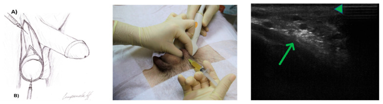
Background and Objectives: Currently, infertility is one of the major problems affecting up to 12% of couples worldwide, with more than a quarter of cases being male-related. It is assumed that Leukocyte-poor platelet-rich plasma (LP-PRP) can improve the function of germ cells and serve as a regenerative substrate as a source of biologically active substances that play an important role in the process of spermatogenesis in infertile men. We aimed to evaluate the proliferation, apoptosis, and growth factors of germ cells after the administration of LP-PRP in patients with non-obstructive azoospermia. Materials and Methods: The study used archival material (paraffin blocks of testicular biopsies) of patients with non-obstructive azoospermia aged 21-34 years (n = 41; associated diagnosis: varicocele). We confirm that no interventions or biopsies were performed as part of the study itself. They were injected bilaterally into the spermatic cord and in the region of the lower pole of the testis under ultrasound control were injected with PRP once a week for 6 weeks. Biopsies were immunohistochemical reactions with antibodies to Ki-67, Bcl-2, caspase 3 and p53, IGF-1, TGF-β, and VEGF-A. Results: Immunohistochemical study of testicular biopsies after LP-PRP injection revealed an increase in the number of cells stained for proliferation proteins (Ki-67) and anti-apoptosis (Bcl-2), IGF-1, TGF-β, VEGF-A; decrease caspase-3- and p53-positive cells. Conclusions: In LP-PRP, platelet α-granule growth factors, which are key regulators of the cell cycle of germ cells, demonstrate restoration of the proliferative-apoptotic balance, confirmed by the expression levels of Ki-67, Bcl-2, caspase 3, and p53 in patients with non-obstructive azoospermia. In human testicular biopsies, the administration of LP-PRP led to an exponential release of numerous growth factors from platelet α-granules, which, based on their regenerative properties, improved the morphological and immunohistochemical picture of the germinal epithelium in non-obstructive azoospermia.
Keywords: PRP; growth factors; non-obstructive azoospermia; spermatogenesis.
Conflict of interest statement
The authors declare no competing financial interests. The authors certify that they have NO affiliations with or involvement in any organization or entity with any financial interest (such as honoraria; educational grants; participation in speakers’ bureaus; membership, employment, consultancies, stock ownership, or other equity interest; and expert testimony or patent-licensing arrangements), or non-financial interest (such as personal or professional relationships, affiliations, knowledge or beliefs) in the subject matter or materials discussed in this manuscript.
Figures


References
-
- Lu J., Liu Z., Shu M., Zhang L., Xia W., Tang L., Li J., Huang B., Li H. Human placental mesenchymal stem cells ameliorate chemotherapy-induced damage in the testis by reducing apoptosis/oxidative stress and promoting autophagy. Stem. Cell Res. Ther. 2021;12:199. doi: 10.1186/s13287-021-02275-z. - DOI - PMC - PubMed
MeSH terms
Substances
Supplementary concepts
LinkOut - more resources
Full Text Sources
Research Materials
Miscellaneous
