

Global Longitudinal Strain in Stress Echocardiography: A Review of Its Diagnostic and Prognostic Role in Noninvasive Cardiac Assessment
- PMID: 40870927
- PMCID: PMC12385223
- DOI: 10.3390/diagnostics15162076
Global Longitudinal Strain in Stress Echocardiography: A Review of Its Diagnostic and Prognostic Role in Noninvasive Cardiac Assessment
Abstract
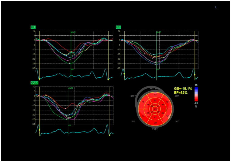
Background: The integration of global longitudinal strain (GLS) with stress echocardiography (SE) represents a significant advancement in non-invasive cardiac diagnostics, particularly in the evaluation of coronary artery disease (CAD). GLS, derived from speckle-tracking echocardiography, quantifies myocardial deformation and offers superior sensitivity for detecting subclinical myocardial dysfunction compared to conventional metrics like wall motion and ejection fraction. Recent studies have validated the prognostic and diagnostic efficacy of GLS both at rest and during stress, notably enhancing the detection of obstructive and non-obstructive CAD, microvascular dysfunction, and other cardiac pathologies. Methods: This manuscript synthesizes extensive clinical data demonstrating the added value of GLS during stress echocardiography across diverse cardiac conditions-including valvular heart disease, heart failure, cardio-oncology, and pediatric cardiology. Novel metrics like longitudinal strain reserve (LSR), myocardial work indices, and post-systolic strain have further enriched risk stratification strategies. Results: The combination of GLS with SE has been shown to approximate the accuracy of invasive coronary angiography in intermediate-risk patients and in cases with equivocal traditional SE findings. Despite its clinical promise, the utility of GLS is challenged by technical limitations, including image quality dependency, inter-vendor variability, and limited applicability during high heart rate states. Conclusions: As technological refinement and standardization progress, GLS integrated with SE is poised to become a mainstay in precision cardiology, improving diagnostic yield, guiding therapeutic decisions, and enhancing patient outcomes.
Keywords: coronary artery disease (CAD); global longitudinal strain (GLS); longitudinal strain reserve (LSR); myocardial deformation; myocardial work; non-invasive cardiac imaging; risk stratification; speckle-tracking echocardiography (STE); stress echocardiography (SE); subclinical dysfunction.
Conflict of interest statement
The authors declare no conflict of interest.
Figures

Similar articles
-
Global longitudinal strain in the prediction of significant coronary artery disease: how accurate is it for patients with a high clinical probability of chronic coronary syndrome and preserved left ventricular ejection fraction?Echo Res Pract. 2025 Jul 1;12(1):16. doi: 10.1186/s44156-025-00084-1. Echo Res Pract. 2025. PMID: 40588772 Free PMC article.
-
Speckle-Tracking Strain Echocardiography for the Assessment of Left Ventricular Structure and Function: A Scientific Statement From the American Heart Association.Circulation. 2025 Aug 6. doi: 10.1161/CIR.0000000000001354. Online ahead of print. Circulation. 2025. PMID: 40765507 Review.
-
Longitudinal Myocardial Deformation as an Emerging Biomarker for Post-Traumatic Cardiac Dysfunction.Life (Basel). 2025 Jun 30;15(7):1052. doi: 10.3390/life15071052. Life (Basel). 2025. PMID: 40724554 Free PMC article. Review.
-
Diagnostic accuracy of left ventricular longitudinal function by speckle tracking echocardiography to predict significant coronary artery stenosis. A systematic review.BMC Med Imaging. 2015 Jul 25;15:25. doi: 10.1186/s12880-015-0067-y. BMC Med Imaging. 2015. PMID: 26204938 Free PMC article.
-
Artificial Intelligence Performance in Cardiac Magnetic Resonance Strain Analysis for Aortic Stenosis: Validation with Echocardiography and Healthy Controls.Medicina (Kaunas). 2025 May 22;61(6):950. doi: 10.3390/medicina61060950. Medicina (Kaunas). 2025. PMID: 40572638 Free PMC article.
References
-
- Woodward W., Dockerill C., McCourt A., Upton R., O’DRiscoll J., Balkhausen K., Chandrasekaran B., Firoozan S., Kardos A., Wong K., et al. Real-world performance and accuracy of stress echocardiography: The EVAREST observational multi-centre study. Eur. Heart J. Cardiovasc. Imaging. 2022;23:689–698. doi: 10.1093/ehjci/jeab092. - DOI - PMC - PubMed
-
- Sicari R., Nihoyannopoulos P., Evangelista A., Kasprzak J., Lancellotti P., Poldermans D., Voigt J.-U., Zamorano J.L., European Association of Echocardiography Stress Echocardiography Expert Consensus Statement—Executive Summary: European Association of Echocardiography (EAE) (a registered branch of the ESC) Eur. Heart J. 2009;30:278–289. doi: 10.1093/eurheartj/ehn492. - DOI - PubMed
-
- Hoffmann R., Lethen H., Marwick T., Arnese M., Fioretti P., Pingitore A., Picano E., Buck T., Erbel R., Flachskampf F.A., et al. Analysis of interinstitutional observer agreement in interpretation of dobutamine stress echocardiograms. J. Am. Coll. Cardiol. 1996;27:330–336. doi: 10.1016/0735-1097(95)00483-1. - DOI - PubMed
-
- Saraste M., Knuuti J., Saraste A. Two-Dimensional Speckle-Tracking during Dobutamine Stress Echocardiography in the Detection of Myocardial Ischemia in Patients with Suspected Coronary Artery Disease. J. Am. Soc. Echocardiogr. 2016;29:470–479.e3. - PubMed
-
- Nagy A.I., Sahlén A., Manouras A., Henareh L., da Silva C., Günyeli E., Apor A.A., Merkely B., Winter R. Combination of contrast-enhanced wall motion analysis and myocardial deformation imaging during dobutamine stress echocardiography. Eur. Heart J. Cardiovasc. Imaging. 2015;16:88–95. doi: 10.1093/ehjci/jeu171. - DOI - PubMed
Publication types
LinkOut - more resources
Full Text Sources
Research Materials
Miscellaneous
