. 2025 Aug 19;15(16):2078.
doi: 10.3390/diagnostics15162078.

Diagnostic and Clinical Outcomes of Three Regenerative Strategies for Alveolar Bone Defects: A Comparative Study Using CBCT and ISQ

Affiliations

Diagnostic and Clinical Outcomes of Three Regenerative Strategies for Alveolar Bone Defects: A Comparative Study Using CBCT and ISQ

Sorin Gheorghe Mihali et al. Diagnostics (Basel). .

Abstract

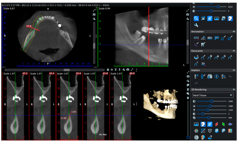
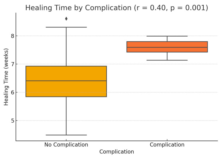
Background: This prospective clinical study aimed to evaluate the effectiveness of platelet-rich fibrin (PRF) in guided bone regeneration (GBR) prior to dental implant placement. Material and methods: Sixty-five patients with alveolar bone defects were randomly assigned to three groups. All groups received a composite graft consisting of 70% allograft and 30% xenograft. Group A received the graft combined with PRF. Group B received the graft with PRF and a resorbable collagen membrane. Group C (control) received the same graft and membrane without PRF. Cone-beam computed tomography (CBCT) was used to assess bone regeneration at baseline and 6 months postoperatively. Implant stability was evaluated using ISQ values at the time of implant placement (6 months after grafting) and again at 3 to 4 months during the second-stage uncovering procedure. Soft tissue healing, postoperative complications, and pain scores were also recorded. Results: Group B showed the best outcomes, with the highest mean vertical bone gain (3.0 ± 0.4 mm), greatest implant stability (ISQ: 74.2 ± 1.8), and no complications. Group A achieved moderate bone gain (2.3 ± 0.4 mm) and good ISQ values (71.5 ± 2.3), with favorable soft tissue healing. In contrast, Group C had the lowest bone gain (2.1 ± 0.5 mm), reduced ISQ values (68.9 ± 2.9), and the highest incidence of complications, including dehiscence and minor infections. Conclusions: These results suggest that PRF enhances both hard and soft tissue regeneration, particularly when used with grafts and membranes. PRF may reduce healing time and postoperative discomfort, improving the overall success of regenerative implant procedures.

Keywords: bone graft; collagen membrane; guided bone regeneration; implant stability; platelet-rich fibrin; soft tissue healing.


Conflict of interest statement

The authors declare no conflicts of interest.

Figures

Figure 1
Figure 1
Study design and treatment workflow.
Figure 2
Figure 2
(a) Autologous PRF membrane prepared on a metal tray before placement. (b) Blend of 30% xenograft (Bio-Oss®, Geistlich Pharma AG, Switzerland) and 70% allograft (Puros Cancellous Particulate Allograft, ZimVie, Germany). (c) Multiple layers of PRF sutured over the defect and stabilized to the periosteum. (d) Final flap closure in Group A using horizontal mattress and single interrupted sutures to ensure stable healing.
Figure 3
Figure 3
(a) Initial clinical view of the bone defect. (b) Placement of the bone graft composed of 30% xenograft (Bio-Oss®, Geistlich Pharma AG, Switzerland) and 70% allograft (Puros Cancellous Particulate Allograft, ZimVie, Germany). (c) Application of a resorbable collagen membrane (Biomed Extend, Zimmer Biomet, Warsaw, IN, USA) over the grafted area. (d) Stabilization of the grafted site using vertical mattress sutures with resorbable material (Chirmax 5-0 DS15, Chirmax, Prague, Czech Republic), prior to final positioning of the PRF membranes.
Figure 4
Figure 4
(a) Final positioning of PRF membranes before flap advancement with resorbable sutures (Chirmax 5-0 DS15, Chirmax, Prague, Czech Republic). (b) Tension-free flap adaptation over the grafted and membrane-covered area. (c) Suturing with a combination of horizontal mattress and single interrupted sutures. (d) Provisional restoration placed to maintain the space and protect the surgical site postoperatively.
Figure 5
Figure 5
(a) A split-thickness flap was elevated by separating the mucosa from the periosteum to allow tension-free repositioning of the soft tissues. (b) The periosteum was incised to release the flap and increase its elasticity, facilitating passive adaptation over the grafted area after augmentation. (c) In this case with dense cortical bone, decortication was performed by creating multiple perforations with a 0.9 mm NTI round bur (Globus, Germany), facilitating medullary bleeding and improving cell recruitment and vascularization. (d) Positioning of the resorbable collagen membrane (Biomed Extend, Zimmer Biomet, Warsaw, IN, USA).
Figure 5
Figure 5
(a) A split-thickness flap was elevated by separating the mucosa from the periosteum to allow tension-free repositioning of the soft tissues. (b) The periosteum was incised to release the flap and increase its elasticity, facilitating passive adaptation over the grafted area after augmentation. (c) In this case with dense cortical bone, decortication was performed by creating multiple perforations with a 0.9 mm NTI round bur (Globus, Germany), facilitating medullary bleeding and improving cell recruitment and vascularization. (d) Positioning of the resorbable collagen membrane (Biomed Extend, Zimmer Biomet, Warsaw, IN, USA).
Figure 6
Figure 6
(a) Application of the bone graft composed of 30% xenograft (Bio-Oss®, Geistlich Pharma AG, Switzerland) and 70% allograft (Puros Cancellous Particulate Allograft, ZimVie, Germany). (b) Compaction of the grafting material into the defect site. (c) Placement of vertical mattress resorbable sutures (Chirmax 5-0 DS15, Chirmax, Prague, Czech Republic) prior to the final positioning of the resorbable collagen membrane (Biomed Extend, Zimmer Biomet, Warsaw, IN, USA). (d) Flap closure was achieved using horizontal mattress and single interrupted sutures with the same resorbable material to ensure tension-free adaptation and stable wound closure.
Figure 7
Figure 7
CBCT-based evaluation at 6 months postoperatively in a representative case. Cross-sectional analysis at 45.6 mm revealed a horizontal ridge width of 11.02 mm and a trabecular bone density estimated at 960 Hounsfield Units (HU). The buccal and lingual cortices were intact, with no evidence of dehiscence or fenestration. The 3D volumetric reconstruction confirmed complete bone coverage and spatial integrity around the radiopaque implant structure.
Figure 8
Figure 8
Comparative analysis of vertical bone gain following regenerative procedures in the three study groups.
Figure 9
Figure 9
Correlation between vertical bone gain and implant stability (ISQ).
Figure 10
Figure 10
Distribution of healing time in patients with and without postoperative complications.
Figure 11
Figure 11
(a) VAS pain scores on postoperative day 1. (b) Healing time in patients with and without complications.
Figure 12
Figure 12
(a) Preoperative slice showing severe horizontal ridge deficiency (width: 1.20 mm). (b) Six-month postoperative slice showing horizontal ridge width increased to 11.63 mm and continuous corticalization, confirming successful bone regeneration and graft mineralization.
Figure 13
Figure 13
Three-dimensional CBCT reconstructions of the posterior mandible. (a) Preoperative rendering showing extensive alveolar resorption and cortical discontinuities. (b) Six-month postoperative reconstruction illustrating volumetric restoration and cortical continuity.
Figure 14
Figure 14
CBCT evaluation of the posterior maxilla at 6 months postoperatively: vertical ridge height (13.79 mm), bucco-palatal width (6.80 mm), and average trabecular density of 685.74 HU, indicative of mature regenerated bone suitable for implant insertion.

Similar articles

References

    1. Urban I.A., Monje A. Guided Bone Regeneration in Alveolar Bone Reconstruction. Oral Maxillofac. Surg. Clin. N. Am. 2019;31:331–338. doi: 10.1016/j.coms.2019.01.003. - DOI - PubMed
    1. Aghaloo T.L., Moy P.K. Which hard tissue augmentation techniques are the most successful in furnishing bony support for implant placement? Int. J. Oral Maxillofac. Implant. 2007;22:49–70. - PubMed
    1. Tolstunov L., Hamrick J.F.E., Broumand V., Shilo D., Rachmiel A. Bone Augmentation Techniques for Horizontal and Vertical Alveolar Ridge Deficiency in Oral Implantology. Oral Maxillofac. Surg. Clin. N. Am. 2019;31:163–191. doi: 10.1016/j.coms.2019.01.005. - DOI - PubMed
    1. Aghaloo T.L., Misch C., Lin G.H., Iacono V.J., Wang H.L. Bone Augmentation of the Edentulous Maxilla for Implant Placement: A Systematic Review. Int. J. Oral Maxillofac. Implant. 2016;31:19–30. doi: 10.11607/jomi.16suppl.g1. - DOI - PubMed
    1. Lin X., Patil S., Gao Y.G., Qian A. The Bone Extracellular Matrix in Bone Formation and Regeneration. Front. Pharmacol. 2020;11:757. doi: 10.3389/fphar.2020.00757. - DOI - PMC - PubMed

LinkOut - more resources