

Severe Aortic Stenosis and Pre-Excitation Syndrome in Pregnancy-A Multidisciplinary Approach
- PMID: 40870950
- PMCID: PMC12385372
- DOI: 10.3390/diagnostics15162099
Severe Aortic Stenosis and Pre-Excitation Syndrome in Pregnancy-A Multidisciplinary Approach
Abstract
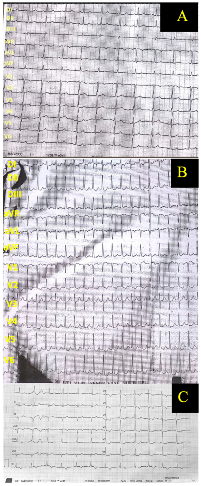
Background/Objectives: Heart disease affects 0.1% to 4% of pregnant women, with congenital heart defects being the leading cause in developed countries. While maternal mortality is generally low, pre-existing cardiac conditions substantially increase adverse outcome risks. This report describes the multidisciplinary management of a pregnant patient with a bicuspid aortic valve, severe aortic stenosis, and ascending aortic ectasia. Case Presentation: A 34-year-old pregnant woman, asymptomatic but at high risk (World Health Organization Class III) for hemodynamic decompensation, was closely monitored throughout gestation. At 36 weeks, intrauterine growth restriction was detected, prompting an elective cesarean delivery at 38 weeks. Postpartum, the patient developed pre-eclampsia, which was managed successfully. Imaging revealed progressive aortic dilation, leading to surgical aortic valve replacement and ascending aorta reduction plasty. Post-operatively, atrioventricular reentrant tachycardia from an unrecognized accessory pathway developed; medical therapy effectively controlled the arrhythmia after failed catheter ablation. One year later, both mother and child remained in good health. Discussion: This case illustrates the complexity of managing pregnancy in women with congenital heart disease and significant aortic pathology. The physiological changes of pregnancy can exacerbate underlying lesions, necessitating individualized risk assessment, vigilant monitoring, and timely intervention. Conclusions: A multidisciplinary approach involving cardiology, obstetrics, anesthesiology, and genetics is essential to optimize outcomes for pregnant women with significant heart disease. As advances in care allow more women with congenital heart defects to reach childbearing age, structured care pathways remain vital for ensuring safe pregnancies and long-term cardiovascular health.
Keywords: aortic stenosis; bicuspid aortic valve; maternal–fetal medicine; pre-eclampsia; pre-excitation syndrome; pregnancy and cardiovascular disease.
Conflict of interest statement
The authors declare no conflicts of interest.
Figures


References
-
- Regitz-Zagrosek V., Roos-Hesselink J.W., Bauersachs J., Blomström-Lundqvist C., Cífková R., De Bonis M., Iung B., Johnson M.R., Kintscher U., Kranke P., et al. 2018 ESC Guidelines for the Management of Cardiovascular Diseases during Pregnancy. Eur. Heart J. 2018;39:3165–3241. doi: 10.1093/eurheartj/ehy340. - DOI - PubMed
-
- Roos-Hesselink J.W., Ruys T.P.E., Stein J.I., Thilén U., Webb G.D., Niwa K., Kaemmerer H., Baumgartner H., Budts W., Maggioni A.P., et al. Outcome of Pregnancy in Patients with Structural or Ischaemic Heart Disease: Results of a Registry of the European Society of Cardiology. Eur. Heart J. 2013;34:657–665. doi: 10.1093/eurheartj/ehs270. - DOI - PubMed
-
- Yap S.-C., Drenthen W., Pieper P.G., Moons P., Mulder B.J.M., Mostert B., Vliegen H.W., van Dijk A.P.J., Meijboom F.J., Steegers E.A.P., et al. Risk of Complications during Pregnancy in Women with Congenital Aortic Stenosis. Int. J. Cardiol. 2008;126:240–246. doi: 10.1016/j.ijcard.2007.03.134. - DOI - PubMed
-
- McKellar S.H., MacDonald R.J., Michelena H.I., Connolly H.M., Sundt T.M., III Frequency of Cardiovascular Events in Women with a Congenitally Bicuspid Aortic Valve in a Single Community and Effect of Pregnancy on Events. Am. J. Cardiol. 2011;107:96–99. doi: 10.1016/j.amjcard.2010.08.061. - DOI - PubMed
Publication types
LinkOut - more resources
Full Text Sources
Research Materials
