Review
. 2025 Aug 21;17(16):2708.
doi: 10.3390/nu17162708.

Type 2 Diabetes and the Multifaceted Gut-X Axes

Affiliations
Review

Type 2 Diabetes and the Multifaceted Gut-X Axes

Hezixian Guo et al. Nutrients. .

Abstract

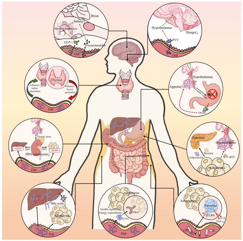
Type 2 diabetes (T2D) is a complex metabolic disease characterized by chronic hyperglycemia due to insulin resistance and inadequate insulin secretion. Beyond the classically implicated organs, emerging evidence highlights the gut as a central player in T2D pathophysiology through its interactions with metabolic organs. The gut hosts trillions of microbes and enteroendocrine cells that influence inflammation, energy homeostasis, and hormone regulation. Disruptions in gut homeostasis (dysbiosis and increased permeability) have been linked to obesity, insulin resistance, and β-cell dysfunction, suggesting multifaceted "Gut-X axes" contribute to T2D development. We aimed to comprehensively review the evidence for gut-mediated crosstalk with the pancreas, endocrine system, liver, and kidneys in T2D. Key molecular mechanisms (incretins, bile acids, short-chain fatty acids, endotoxins, etc.) were examined to construct an integrated model of how gut-derived signals modulate metabolic and inflammatory pathways across organs. We also discuss clinical implications of targeting Gut-X axes and identify knowledge gaps and future research directions. A literature search (2015-2025) was conducted in PubMed, Scopus, and Web of Science, following PRISMA guidelines (Preferred Reporting Items for Systematic Reviews). Over 150 high-impact publications (original research and review articles from Nature, Cell, Gut, Diabetologia, Lancet Diabetes & Endocrinology, etc.) were screened. Data on gut microbiota, enteroendocrine hormones, inflammatory mediators, and organ-specific outcomes in T2D were extracted. The GRADE framework was used informally to prioritize high-quality evidence (e.g., human trials and meta-analyses) in formulating conclusions. T2D involves perturbations in multiple Gut-X axes. This review first outlines gut homeostasis and T2D pathogenesis, then dissects each axis: (1) Gut-Pancreas Axis: how incretin hormones (GLP-1 and GIP) and microbial metabolites affect insulin/glucagon secretion and β-cell health; (2) Gut-Endocrine Axis: enteroendocrine signals (e.g., PYY and ghrelin) and neural pathways that link the gut with appetite regulation, adipose tissue, and systemic metabolism; (3) Gut-Liver Axis: the role of microbiota-modified bile acids (FXR/TGR5 pathways) and bacterial endotoxins in non-alcoholic fatty liver disease (NAFLD) and hepatic insulin resistance; (4) Gut-Kidney Axis: how gut-derived toxins and nutrient handling intersect with diabetic kidney disease and how incretin-based and SGLT2 inhibitor therapies leverage gut-kidney communication. Shared mechanisms (microbial SCFAs improving insulin sensitivity, LPS driving inflammation via TLR4, and aryl hydrocarbon receptor ligands modulating immunity) are synthesized into a unified model. An integrated understanding of Gut-X axes reveals new opportunities for treating and preventing T2D. Modulating the gut microbiome and its metabolites (through diet, pharmaceuticals, or microbiota therapies) can improve glycemic control and ameliorate complications by simultaneously influencing pancreatic islet function, hepatic metabolism, and systemic inflammation. However, translating these insights into clinical practice requires addressing gaps with robust human studies. This review provides a state-of-the-art synthesis for researchers and clinicians, underlining the gut as a nexus for multi-organ metabolic regulation in T2D and a fertile target for next-generation therapies.

Keywords: NAFLD; bile acids; gut microbiome; gut–liver axis; incretin hormones; metabolic inflammation; short-chain fatty acids; type 2 diabetes.


Conflict of interest statement

The authors declare that they have no competing interests.

Figures

Figure 1
Figure 1
Gut–Pancreas Axis. Dietary glucose stimulates intestinal cells to secrete GLP-1 and GIP. A balanced microbiota (green microbes) produces short-chain fatty acids (SCFAs), which enhance the intestinal barrier. Conversely, a dysbiotic microbiota (red microbes) generates LPS and diabetic metabolites (e.g., TMAO and BCAAs), impairing β-cell function and insulin action. Substances such as GIP, GLP-1, BCAAs, LPS, and TMAO reach the pancreas via the vagus nerve, influencing insulin and glucagon secretion in a glucose-dependent manner. Green arrows represent protective pathways for metabolism and inflammation. Red arrows represent damaging pathways.
Figure 2
Figure 2
Gut–Endocrine Axis: Gut–brain signaling and interactions among the intestine, adipose tissue, and other hormone-secreting organs. The intestine and brain are intimately connected via neural and endocrine pathways. After feeding, GLP-1 and PYY is secreted by L-cells, and oxyntomodulin is secreted by the intestine, clarifying that GLP-1’s effect on the arcuate nucleus (ARC) is indirect. We note that GLP-1 acts via the median eminence (a BBB-free zone adjacent to the ARC) and possibly through tanycytes or other intermediary cells, rather than directly entering the ARC. However, physiologically, oxyntomodulin levels are low—only after bariatric surgery do its concentrations rise enough to have significant effects—so its normal role in appetite regulation remains unclear. During fasting, gastric ghrelin stimulates the same brain regions to enhance appetite. The intestine communicates with adipose tissue by regulating adipokine release and systemic lipid metabolism. Bile acids, SCFAs, and LPS have been suggested to modulate adipogenesis, energy expenditure, and inflammation (e.g., the browning of adipose tissue and activation of macrophage TLR4 pathways), though direct evidence in humans is limited and comes mainly from animal studies. Intestinal LPS infiltrates adipose tissue to activate TLR4 in adipose macrophages, inducing the production of IL-6 and TNF-α. SCFAs produced by gut microbiota bind to GPR43 receptors on adipocytes, inhibiting insulin signaling, restricting fat storage, and promoting energy expenditure. The intestine microbiota influences cortisol activity by affecting 11β-HSD in the liver and adipose tissue; the hypothalamus releases chronic stress hormones to inversely affect the kidneys’ regulation of the HPA, thereby promoting intestinal dysbiosis. The intestine–thyroid axis refers to the process where intestinal bacteria promote thyroid hormones to bind with the thyroid gland and affect iodine uptake, while hypothyroidism can, in turn, slow down intestinal peristalsis and influence the microbiota. Solid arrows denote hormone/metabolite transport via the bloodstream. Dashed arrows denote neural conduction. Green arrows represent protective metabolic and anti-inflammatory pathways. Red arrows represent damaging pathways.
Figure 3
Figure 3
Gut–Liver Axis. Hepatocytes synthesize primary bile acids from cholesterol, which are stored in the gallbladder and released into the small intestine during meals. Bile acids activate FXR in the intestine, prompting the secretion of FGF19. FGF19 travels via the portal vein to the liver, where it inhibits CYP7A1, thereby negatively regulating de novo bile acid synthesis and reducing hepatic gluconeogenesis. FXR signaling also improves insulin sensitivity and suppresses hepatic lipid synthesis. Gut microbiota convert primary bile acids to secondary bile acids. Secondary bile acids DCA/LCA can not only activate TGR5 receptor on L-cells to promote GLP-1 secretion but also activate TGR5 receptor on Kupffer cells to increase cAMP, thereby inhibiting TNF-α/IL-1β and alleviating liver inflammation. High-fat diets induce intestinal leakage, allowing LPS to trigger the TLR4-NF-κB pathway in Kupffer cells, leading to proinflammatory cytokine release. Gut microbes produce SCFAs to regulate insulin sensitivity and gluconeogenesis; intestinal-derived ethanol exacerbates oxidative stress and lipid peroxidation, while TMA/TMAO promotes atherosclerosis and disrupts cholesterol efflux. Green arrows denote protective metabolic and inflammatory pathways; red arrows indicate damaging pathways.
Figure 4
Figure 4
Gut–Kidney Axis: Three phases of the gut–kidney axis in type 2 diabetes (T2D), chronic kidney disease (CKD), and diabetic kidney disease (DKD). Beneficial bacteria (green microbes) produce SCFAs and IPA, while intestinal L-cells secrete GLP-1, collectively maintaining barrier function and exerting anti-inflammatory effects. IPA reduces IL-6 production by activating the PXR/AhR pathway. Harmful bacteria (red microbes) metabolize uremic toxin precursors (e.g., from p-cresol to pCS, indole to IS, TMA to TMAO, and PAA to PAG). The uremic toxin pCS enters renal tubular epithelial cells via an organic anion transporter (OAT), inducing NADPH oxidase to generate ROSs and activate inflammatory signals, such as NF-κB and NLRP3. IS exacerbates inflammation through the AhR-NF-κB-P450 pathway, while TMAO activates NF-κB to promote IL-6 and NLRP3 release. Reduced renal function allows urea and uremic toxins to diffuse into the intestine, where urease converts them to ammonia, increasing the luminal pH, disrupting the intestinal barrier, and promoting dysbiosis. Current therapeutic strategies include GLP-1 RAs and SGLT2i. Green arrows represent protective metabolic and inflammatory pathways; red arrows denote damaging pathways.
Figure 5
Figure 5
Integrative Model of Gut-X Axes in T2D. A multi-organ metabolic regulatory network centered on the gut microbiota, which systematically demonstrates that dietary fiber and high-fat diets coordinately regulate glucose–lipid metabolism, inflammatory responses, and insulin sensitivity across multiple organs by influencing microbial metabolites, immune signals, and hormonal pathways, revealing their key roles in the pathogenesis of T2D. Many pathways illustrated (e.g., microbial metabolite effects on the host’s metabolism) are hypothetical or proposed and remain to be confirmed, especially in humans. The figure shows that dietary fiber can promote intestinal microbes to produce SCFAs and AhR, while a high-fat diet promotes LPS production in the intestine. (A) Dietary fiber is fermented in the colon to short-chain fatty acids (SCFAs: acetate, propionate, butyrate), which act via FFAR2/3 and HDAC inhibition to stimulate L-cell secretion of GLP-1 and PYY, enhancing satiety and insulin secretion; expand Tregs and strengthen the intestinal barrier to dampen metabolic inflammation; and, via the portal vein, act on the liver to suppress gluconeogenesis and lipogenesis, improving insulin sensitivity. (B) Fermentation products of fiber or tryptophan metabolites activate AhR signaling, promoting ILC3/Th17-derived IL-22 to repair and fortify the barrier, thereby reducing LPS flux to the liver; hepatic fatty acid oxidation increases and lipid accumulation decreases, lowering NAFLD risk. (C) A high-fat diet disrupts the barrier and drives metabolic endotoxemia (LPS-TLR4), activating NF-κB and upregulating TNF-α/IL-6; overflow of free fatty acids (FFA) induces insulin resistance in liver, muscle, and adipose tissue, exacerbates β-cell stress and apoptosis, and leads to hepatic steatosis (NAFLD); central appetite and leptin signaling become dysregulated, further worsening the metabolic phenotype. (D) BCAAs, endocannabinoids (eCBs), and gut hormones (GLP-1, PYY) jointly regulate muscle metabolic stress, pancreatic β-cell function, and whole-body energy homeostasis; their imbalance can amplify insulin resistance and fatty liver.Green arrows signify beneficial pathways (metabolic improvement, anti-inflammation, and barrier enhancement); red arrows indicate harmful signals (proinflammation, insulin resistance, and fat accumulation).

Similar articles

References

    1. Ong K.L., Stafford L.K., McLaughlin S.A., Boyko E.J., Vollset S.E., Smith A.E., Dalton B.E., Duprey J., Cruz J.A., Hagins H., et al. Global, Regional, and National Burden of Diabetes from 1990 to 2021, with Projections of Prevalence to 2050: A Systematic Analysis for the Global Burden of Disease Study 2021. Lancet. 2023;402:203–234. doi: 10.1016/S0140-6736(23)01301-6. - DOI - PMC - PubMed
    1. Dludla P.V., Mabhida S.E., Ziqubu K., Nkambule B.B., Mazibuko-Mbeje S.E., Hanser S., Basson A.K., Pheiffer C., Kengne A.P. Pancreatic β-Cell Dysfunction in Type 2 Diabetes: Implications of Inflammation and Oxidative Stress. World J. Diabetes. 2023;14:130–146. doi: 10.4239/wjd.v14.i3.130. - DOI - PMC - PubMed
    1. Badkas A., Pacheco M.P., Sauter T. Network Modeling Approaches for Metabolic Diseases and Diabetes. Curr. Opin. Syst. Biol. 2024;39:100530. doi: 10.1016/j.coisb.2024.100530. - DOI
    1. Phatak S.R., Saboo B., Dwivedi S., Zinzuwadia P., Panchal D., Ganguli A., Hasnani D. Sweetening Sixteen: Beyond the Ominous Octet. J. Diabetol. 2021;12:1–9. doi: 10.4103/jod.jod_9_20. - DOI
    1. Baars D.P., Fondevila M.F., Meijnikman A.S., Nieuwdorp M. The Central Role of the Gut Microbiota in the Pathophysiology and Management of Type 2 Diabetes. Cell Host Microbe. 2024;32:1280–1300. doi: 10.1016/j.chom.2024.07.017. - DOI - PubMed

MeSH terms

LinkOut - more resources