. 2025 Aug 19;14(8):820.
doi: 10.3390/pathogens14080820.

Antiviral Activity of Medicinal Plant Extracts Vitex negundo and Macaranga tanarius Against SARS-CoV-2

Affiliations

Antiviral Activity of Medicinal Plant Extracts Vitex negundo and Macaranga tanarius Against SARS-CoV-2

Muhareva Raekiansyah et al. Pathogens. .

Abstract

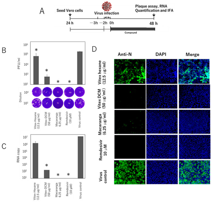
Natural products possess a wide range of biological and biochemical potentials, with plant-derived compounds being significant sources for discovering new drugs. In this study, extracts of Vitex negundo and Macaranga tanarius prepared with different solvents were tested for their antiviral activity against the original SARS-CoV-2 Wuhan strain and its variants using plaque assay, quantitative real time RT-PCR, and immunofluorescence assay (IFA). Our results showed that at their maximum non-toxic concentrations, Vitex-Dichloromethane (DCM) and Macaranga extracts significantly inhibited SARS-CoV-2 Wuhan strain growth in Vero E6 cells, showing a 5-log reduction in plaque assay and confirmed by IFA. Meanwhile, Vitex-Hexane showed moderate activity with a 2-log decrease. The inhibition was shown in a dose-dependent manner. The antiviral efficacy of these extracts was further demonstrated against various SARS-CoV-2 variants including Alpha, Beta, Delta, and Omicron. Both Vitex-DCM and Macaranga showed strong virucidal activity. In addition, Vitex-DCM and Macaranga inhibited the transcriptional activity of purified SARS-CoV-2 RdRp, indicating that RdRp inhibition may contribute to viral suppression as shown at the post-infection stage. Furthermore, combining Vitex-DCM or Macaranga with remdesivir showed a synergistic effect against SARS-CoV-2. These results suggest that Vitex negundo and Macaranga tanarius extracts are promising candidates for anti-SARS-CoV-2 treatments. Their synergy with remdesivir also underscores the potential of drug combinations in fighting SARS-CoV-2 and preventing the emergence of mutant variants.

Keywords: Macaranga tanarius; SARS-CoV-2; Vitex negundo; antiviral.


Conflict of interest statement

The authors declare no conflicts of interest.

Figures

Figure 1
Figure 1
Schematic representation of the workflow for in vitro drug testing of plant extracts against SARS-CoV-2 using the Vero-E6 cell model: (1) plant extracts were prepared using different solvents, (2) confluent Vero-E6 cells were infected with SARS-CoV-2 in the presence of the samples, (3) the cells were incubated for 48 h, (4) viral inhibition was measured using various methods, including plaque assay, RT-qPCR, and immunofluorescence assay (IFA), and (5) data were analyzed and interpreted.
Figure 2
Figure 2
Cell viability assay. Vero cells E6 in 96-well plates were treated with Extracts-Hexane (EH), Extract-DCM, or Extract-M (A) and remdesivir (B) with increasing concentration. After 48 h incubation, cell viability was determined by MTT assay as described in the “Materials and Methods” section. The results are presented as the mean (±SD) of two independent experiments.
Figure 3
Figure 3
Evaluation of the antiviral activity in the (1) Vitex-Hexane extracts, (2) Vitex-DCM extracts, and (3) Macaranga extracts against SARS-CoV-2, assessed by using standard plaque assays and Vero E6 cells. Cells were treated with extracts at their maximum non-toxic concentrations. Remdesivir was used as a positive control. After 48 h, the level of infectious virus in the culture fluids was determined. The upper panel shows viral titers and cell viability; the lower panel shows representative plaques. Data are mean ± SD from two independent experiments. *: A p-value of <0.05 vs. the control is considered statistically significant.
Figure 4
Figure 4
Antiviral activity of Extract-Hexane, Extract-DCM, and Extract-M against SARS-CoV-2 was evaluated using an immunofluorescence assay. Vero cells were infected with the Wuhan strain (MOI 0.05) and treated with extracts at the indicated concentrations. Remdesivir served as a positive control. After 48 h, cells were stained with anti-nucleocapsid antibody (green) and DAPI (blue) for fluorescence microscopy.
Figure 5
Figure 5
Antiviral activity of the extracts against SARS-CoV-2 variants. Vero E6 cells were infected (MOI 0.05) with Alpha (A), Beta (B), Delta (C), or Omicron BA.1 (D) variants and treated with the extracts at indicated concentrations. Remdesivir was used as a control. After 48 h, the infectious virus in the culture fluids was measured by plaque assays. Data are shown as mean ± SD of two independent experiments virus titers in the infectious culture fluid were measured by plaque assay. Data are shown as mean ± SD of two independent experiments. *: A p-value of <0.05 vs. the control is considered statistically significant.
Figure 6
Figure 6
Time-of-addition assay (virus pre-treatment). (A) Schematic diagram of the viral inhibition assay. SARS-CoV-2 (MOI 0.05) was mixed with extracts or remdesivir at the indicated concentrations for 1 h and added to Vero E6 cells. After 48 h inoculation, viral inhibition was assessed using (B) plaque assay showing viral titers (top) and representative plaques (bottom), (C) viral RNA levels by RT-qPCR, and (D) immunofluorescence staining targeting the nucleocapsid protein. Untreated infected cells served as infection controls. Data are shown as mean ± SD from two independent experiments. *: A p-value of <0.05 vs. the control is considered statistically significant.
Figure 7
Figure 7
Time-of-addition assay (post-entry treatment). Vero E6 cells were infected with SARS-CoV-2 (MOI 0.05) for 1 h. After virus adsorption, the inoculum was removed, cells were washed, and fresh medium containing extracts or remdesivir was added. After 48 h, viral inhibition was evaluated. (A) Schematic of the post-entry treatment setup, (B) plaque assay showing viral titers (top) and representative plaques (bottom), (C) RT-qPCR for viral RNA levels, and (D) immunofluorescence staining for nucleocapsid protein. Data are shown as mean ± SD from two independent experiments. *: A p-value of <0.05 vs. the control is considered statistically significant.
Figure 8
Figure 8
Inhibition of SARS-CoV-2 RNA-dependent RNA polymerase (RdRp) by Vitex-DCM and Macaranga. The inhibitory effects of Vitex-DCM at concentration of 200 μg/mL and 100 μg/mL and Macaranga at concentration of 50 μg/mL and 25 μg/mL were examined using an in vitro enzyme-based RdRp reporter assay. Purified RdRp complexes were incubated with varying concentration of the sample extracts and the resulting polymerization activity was quantified using fluorescent plate reader. The transcriptional activity of control samples treated with 0.1% DMSO was considered 0% for calculating relative enzymatic inhibition, while 100% inhibition was determined using positive control (10 µM Remdesivir). The results shown are the mean (±SD) of two independent experiments.
Figure 9
Figure 9
Synergistic effect of Vitex-DCM or Macaranga with remdesivir. Vero E6 cells were exposed to the SARS-CoV-2 Wuhan strain at a multiplicity of infection (MOI) of 0.05 in the presence of a combination of extract samples and remdesivir at the indicated concentrations. (A) Viral titers measured by plaque assay on Vero-TM cells and (B) representative plaques for the combination of Vitex-DCM and remdesivir. (C) Viral titers by plaque assay and (D) representative plaques of the combination of Macaranga and remdesivir. The results are presented as the mean (±SD) of two independent experiments. *: A p-value of <0.05 vs. the control is considered statistically significant.

References

    1. Fouchier R.A., Kuiken T., Schutten M., van Amerongen G., van Doornum G.J., van den Hoogen B.G., Peiris M., Lim W., Stöhr K., Osterhaus A.D. Aetiology: Koch’s postulates fulfilled for SARS virus. Nature. 2003;423:240. doi: 10.1038/423240a. - DOI - PMC - PubMed
    1. Lu H., Stratton C.W., Tang Y.W. Outbreak of pneumonia of unknown etiology in Wuhan, China: The mystery and the miracle. J. Med. Virol. 2020;92:401–402. doi: 10.1002/jmv.25678. - DOI - PMC - PubMed
    1. Naseer S., Khalid S., Parveen S., Abbass K., Song H., Achim M.V. COVID-19 outbreak: Impact on global economy. Front. Public Health. 2023;10:1009393. doi: 10.3389/fpubh.2022.1009393. - DOI - PMC - PubMed
    1. Huang Y.Z., Kuan C.C. Vaccination to reduce severe COVID-19 and mortality in COVID-19 patients: A systematic review and meta-analysis. Eur. Rev. Med. Pharmacol. Sci. 2022;26:1770–1776. doi: 10.26355/eurrev_202203_28248. - DOI - PubMed
    1. Flacco M.E., Acuti Martellucci C., Baccolini V., De Vito C., Renzi E., Villari P., Manzoli L. COVID-19 vaccines reduce the risk of SARS-CoV-2 reinfection and hospitalization: Meta-analysis. Front. Med. 2022;9:1023507. doi: 10.3389/fmed.2022.1023507. - DOI - PMC - PubMed

LinkOut - more resources