. 2025 Jul 31;18(8):1145.
doi: 10.3390/ph18081145.

Screening of an FDA-Approved Drug Library: Menadione Induces Multiple Forms of Programmed Cell Death in Colorectal Cancer Cells via MAPK8 Cascades

Affiliations

Screening of an FDA-Approved Drug Library: Menadione Induces Multiple Forms of Programmed Cell Death in Colorectal Cancer Cells via MAPK8 Cascades

Liyuan Cao et al. Pharmaceuticals (Basel). .

Abstract

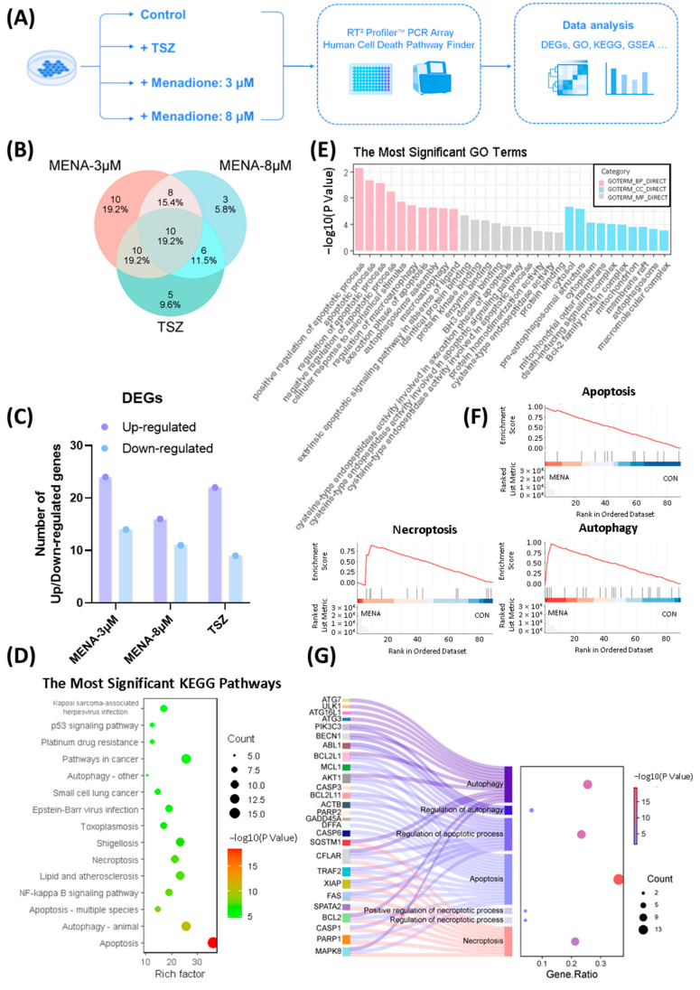
Background: Colorectal cancer (CRC) is a prevalent gastrointestinal malignancy, ranking third in incidence and second in cancer-related mortality. Despite therapeutic advances, challenges such as chemotherapy toxicity and drug resistance persist. Thus, there is an urgent need for novel CRC treatments. However, developing new drugs is time-consuming and resource-intensive. As a more efficient approach, drug repurposing offers a promising alternative for discovering new therapies. Methods: In this study, we screened 1068 small molecular compounds from an FDA-approved drug library in CRC cells. Menadione was selected for further study based on its activity profile. Mechanistic analysis included a cell death pathway PCR array, differential gene expression, enrichment, and network analysis. Gene expressions were validated by RT-qPCR. Results: We identified menadione as a potent anti-tumor drug. Menadione induced three programmed cell death (PCD) signaling pathways: necroptosis, apoptosis, and autophagy. Furthermore, we found that the anti-tumor effect induced by menadione in CRC cells was mediated through a key gene: MAPK8. Conclusions: By employing methods of cell biology, molecular biology, and bioinformatics, we conclude that menadione can induce multiple forms of PCD in CRC cells by activating MAPK8, providing a foundation for repurposing the "new use" of the "old drug" menadione in CRC treatment.

Keywords: MAPK8; colorectal cancer; drug screening; menadione; necroptosis; programmed cell death.


Conflict of interest statement

The authors declare no conflicts of interest.

Figures

Figure 1
Figure 1
Screening of an FDA-approved drug library to identify effective compounds of inducing cell necroptosis. (A) Flow chart of drug screening to identify potential anti-tumor drugs that could induce necroptotic cell death. 1068 drugs in an FDA-approved drug library were screened, and menadione was identified as a promising candidate. (B) Cell viability of L929-FADD-KO cells after treating with 12 candidate drugs (10 μM, 24 h) that caused >50% reduction in cell viability (n = 3). (C) Classification of 12 candidate drugs. (D) Chemical structure of menadione. (E,F) Nec-1 (30 μM) was used to rescue cell death induced by candidate drugs (10 μM, 24 h) in L929-FADD-KO cells (E) and wild-type L929 (F). Statistical significance was evaluated using unpaired two-tailed Student’s t-test. Data of cell viability are presented as mean ± standard error of the mean (SEM, n = 3). * p < 0.05, ** p < 0.01, *** p < 0.001.
Figure 2
Figure 2
Menadione exerted strong anti-tumor effects on colorectal cancer cell lines. (A) Different concentrations of TNF-α were administered to HT-29 cells combined with Smac mimetics (100 nM) and Z-VAD-FMK (20 μM). Cell viability was assessed after 24 h (n = 3). (B) Different concentrations of menadione were administered to HT-29 cells and cell viability was assessed after 24 h (n = 3). (C) Different concentrations of TNF-α were administered to SW620 cells combined with Smac mimetics (100 nM) and Z-VAD-FMK (20 μM). Cell viability was assessed after 24 h(n = 3). (D) Different concentrations of menadione were administered to SW620 cells and cell viability was assessed after 24 h (n = 3). S: Smac mimetics; Z: Z-VAD-FMK. Data of cell viability are presented as mean ± SEM (n = 3).
Figure 2
Figure 2
Menadione exerted strong anti-tumor effects on colorectal cancer cell lines. (A) Different concentrations of TNF-α were administered to HT-29 cells combined with Smac mimetics (100 nM) and Z-VAD-FMK (20 μM). Cell viability was assessed after 24 h (n = 3). (B) Different concentrations of menadione were administered to HT-29 cells and cell viability was assessed after 24 h (n = 3). (C) Different concentrations of TNF-α were administered to SW620 cells combined with Smac mimetics (100 nM) and Z-VAD-FMK (20 μM). Cell viability was assessed after 24 h(n = 3). (D) Different concentrations of menadione were administered to SW620 cells and cell viability was assessed after 24 h (n = 3). S: Smac mimetics; Z: Z-VAD-FMK. Data of cell viability are presented as mean ± SEM (n = 3).
Figure 3
Figure 3
Necroptosis was not the only form of cell death induced by menadione in colorectal cancer cells. (A,B) Flow charts of time-course and concentration–gradient experiments. (C) Menadione (8 μM) or TSZ (T: TNF-α, 10 ng/mL; S: Smac mimetics, 100 nM; Z: Z-VAD-FMK, 20 μM) was administered to HT-29 cells and cell viability was assessed at indicated time points (0.5 h, 2 h, 5 h, 8 h, and 24 h) (n = 3). Some error bars too small to display clearly. (D) Menadione of different doses was added to HT-29 cells and cell viability was determined 1 h after treatment (n = 3). Some error bars too small to display clearly. (E) Flow chart of Nec-1 rescue assay. (F) Menadione (1 μM, 3 μM, 6 μM and 8 μM) was administered to HT-29 cells with or without Nec-1 (10 μM) and cell viability was assessed after 1 h (n = 3) (G) TSZ (T: TNF-α, 10 ng/mL; S: Smac mimetics, 100 nM; Z: Z-VAD-FMK, 20 μM) was administered to HT-29 cells with or without Nec-1 (10 μM) and cell viability was assessed after 24 h (n = 3). Statistical significance was evaluated using an unpaired two-tailed Student’s t-test. Data of cell viability are presented as mean ± SEM (n = 3). ** p < 0.01, *** p < 0.001.
Figure 4
Figure 4
Menadione induced not only necroptosis, but also apoptosis and autophagy in colorectal cancer cells. (A) Flow chart of PCR Array assay and study design. (B,C) Differential analysis was performed and DEGs of experimental groups (MENA-3 μM, MENA-8 μM, and TSZ) were summarized in Venn diagram (B) and histogram (C). (D,E) Enrichment analysis was performed on menadione groups, including KEGG pathway analysis (D) and GO enrichment analysis (E). The most significant KEGG pathways (Top 15) were summarized in (D) and the most significant GO pathways (Top 10 of BP, CC, and MF) were summarized in (E). (F) Representative GSEA pathways: necroptosis, apoptosis and autophagy. GSEA was performed based on the average log2 FC of MENA-3 μM and MENA-8 μM treatment groups relative to the control group, to identify pathways consistently regulated across both conditions. (G) Interactive analysis of necroptosis, apoptosis and autophagy pathways. The Sankey diagram on the left displays genes (e.g., ATG7, CFLAR, BCL2, and MAPK8) mapped to biological processes of necroptosis, apoptosis and autophagy. The bubble plot on the right indicates enrichment degree and statistical significance of each pathway.
Figure 5
Figure 5
The anti-tumor effect induced by menadione in colorectal cancer cells was mediated through MAPK8. (A) Number of up/down-regulated DEGs in pathways of apoptosis, necroptosis, and autophagy after menadione treatment. (B) Venn diagram of DEGs in pathways of apoptosis, necroptosis, and autophagy after menadione treatment. (C) Core hub genes of menadione groups. Cytohubba was used to predict hub genes of 47 DEGs. The MCC algorithm was applied, and top 10 hub genes were displayed. (D) PPI network of DEGs after menadione treatment, visualized by Cytoscape. The nodes in red represent up-regulated DEGs after menadione treatment, and nodes in blue indicate down-regulated DEGs. The node size represents the extent of the gene’s interaction with other genes, with larger nodes indicating a greater number of interacting genes. The edge thickness represents the combined score between two genes, with thicker lines indicating a higher score. The boxed region emphasizes MAPK8 as a core gene in the network. (E) Heatmap of DEGs among menadione and TSZ groups. The color of the heatmap squares represents gene expression level, with darker colors indicating a higher gene expression level. (F) GO and KEGG pathways involving the hub gene MAPK8. Node size and node color represent the number of enriched DEGs in each pathway, with larger sizes and darker colors indicating a higher number of DEGs. (G) PPI network of DEGs that participate in necroptosis, apoptosis, and autophagy pathways. The node color represents the pathways each gene participates in, and the edge indicates protein interaction between two genes.
Figure 6
Figure 6
Validation of programmed cell death and upregulation of MAPK8 cascades induced by menadione in colorectal cancer cells (AD). Relative transcriptional expression level of PCD-related genes (necroptosis, apoptosis, and autophagy) after menadione application was measured by RT-qPCR (n = 3). Menadione: 3 μM or 8 μM; TSZ: TNF-α, 10 ng/mL; Smac mimetics, 100 nM; Z-VAD-FMK, 20 μM. (EG) The relative mRNA levels of MAPK8 were measured by RT-qPCR following treatment with menadione (8 μM) in HT-29, SW620, and HCT116 cells (n = 3). (H) A schematic diagram of the mechanism by which menadione induces multiple forms of PCD. Data are presented as mean ± SEM (n = 3). * p < 0.05, ** p < 0.01, *** p < 0.001.
Figure 7
Figure 7
Drug targets of Menadione and the role of MAPK8 in CRC. (A) PPI analysis of the 33 drug targets was performed using the STRING database, setting a threshold of PPI score > 0.400. Visualization was performed using Cytoscape, where node size and color intensity represent the degree of interaction with other genes. Larger nodes and darker colors indicate a higher number of interacting genes. (B) The DisGeNET database was utilized to retrieve disease targets associated with CRC (search term: colorectal cancer, C1527249), selecting those with a score > 0.3, resulting in a total of 264 targets. A PPI analysis was performed between these 264 CRC-related genes and 33 drug targets of menadione, with a threshold set at PPI score > 0.950. Visualization was performed using Cytoscape, where blue nodes represent the drug targets of menadione and red nodes the CRC disease targets. (C) PPI analysis of MAPK8 and menadione drug targets was conducted using the STRING database, with a threshold set as PPI score > 0.400. Visualization was performed using Cytoscape software, where the size and color intensity of the nodes indicate the degree of interactions with other genes. Larger nodes and darker colors reflect a greater number of interacting genes. (D) Analysis of MAPK8 in conjunction with 264 CRC-related genes was performed using the STRING database, with a threshold set as PPI score > 0.700. This analysis identified 30 genes that exhibit interactions with MAPK8. Visualization was performed using Cytoscape, where the size and color intensity of the nodes represent the degree of interactions with other genes. Larger nodes and darker colors indicate a greater number of interacting genes. (E) Expression level of MAPK8 in CRC patients at different stages (GEPIA database). (F) Kaplan–Meier survival analysis of MAPK8 (229664_at) expression in CRC patients. Patients were stratified into high-expression (n = 852) and low-expression (n = 315) groups based on the optimal cutoff value automatically determined by the Kaplan–Meier Plotter tool.

Similar articles

References

    1. Siegel R.L., Wagle N.S., Cercek A., Smith R.A., Jemal A. Colorectal cancer statistics, 2023. CA Cancer J. Clin. 2023;73:233–254. doi: 10.3322/caac.21772. - DOI - PubMed
    1. Dekker E., Tanis P.J., Vleugels J.L.A., Kasi P.M., Wallace M.B. Colorectal cancer. Lancet. 2019;394:1467–1480. doi: 10.1016/S0140-6736(19)32319-0. - DOI - PubMed
    1. Masoomi H., Ziogas A., Lin B.S., Barleben A., Mills S., Stamos M.J., Zell J.A. Population-based evaluation of adenosquamous carcinoma of the colon and rectum. Dis. Colon. Rectum. 2012;55:509–514. doi: 10.1097/DCR.0b013e3182420953. - DOI - PMC - PubMed
    1. Menon G., Cagir B. StatPearls. StatPearls Publishing LLC.; Treasure Island, FL, USA: 2025. Colon Cancer. - PubMed
    1. Kuipers E.J., Grady W.M., Lieberman D., Seufferlein T., Sung J.J., Boelens P.G., van de Velde C.J., Watanabe T. Colorectal cancer. Nat. Rev. Dis. Primers. 2015;1:15065. doi: 10.1038/nrdp.2015.65. - DOI - PMC - PubMed

LinkOut - more resources