

Pathology, Tissue Distribution, and Phylogenetic Characterization of Largemouth Bass Virus Isolated from a Wild Smallmouth Bass (Micropterus dolomieu)
- PMID: 40872746
- PMCID: PMC12390719
- DOI: 10.3390/v17081031
Pathology, Tissue Distribution, and Phylogenetic Characterization of Largemouth Bass Virus Isolated from a Wild Smallmouth Bass (Micropterus dolomieu)
Abstract
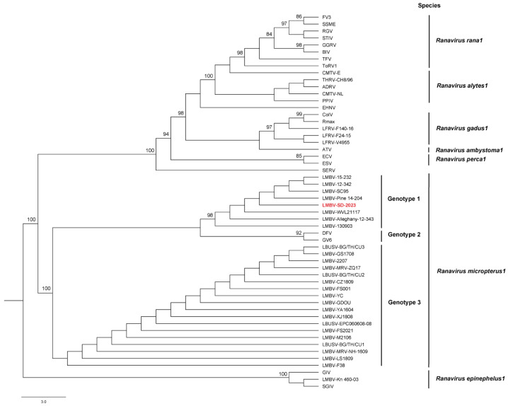
We performed a diagnostic disease investigation on a wild smallmouth bass (Micropterus dolomieu) with skin ulcers that was collected from Lake Oahe, South Dakota, following reports from anglers of multiple fish with similar lesions. Gross and histologic lesions of ulcerative dermatitis, myositis, and lymphocytolysis within the spleen and kidneys were consistent with largemouth bass virus (LMBV) infection. LMBV was detected by conventional PCR in samples of a skin ulcer, and the complete genome sequence of the LMBV (99,184 bp) was determined from a virus isolate obtained from a homogenized skin sample. A maximum likelihood (ML) phylogenetic analysis based on the major capsid protein (MCP) gene alignment supported the LMBV isolate (LMBV-SD-2023) as a member of the species Ranavirus micropterus1, branching within the subclade of LMBV isolates recovered from North American largemouth (Micropterus salmoides) and smallmouth bass. This is the first detection of LMBV in wild smallmouth bass from South Dakota. The ultrastructure of the LMBV isolate exhibited the expected icosahedral shape of virions budding from cellular membranes. Viral nucleic acid in infected cells was visualized via in situ hybridization (ISH) within dermal granulomas, localized predominantly at the margin of epithelioid macrophages and central necrosis. Further sampling is needed to determine the geographic distribution, affected populations, and evolutionary relationship between isolates of LMBV.
Keywords: iridovirus; largemouth bass virus; ranavirus; smallmouth bass.
Conflict of interest statement
The authors declare that there are no competing conflicts of interest related to this work. Any use of trade, firm, or product names is for descriptive purposes only and does not imply endorsement by state or the U.S. Government.
Figures


Similar articles
-
Phylogenomic Characterization of Ranavirus Isolated from Wild Smallmouth Bass (Micropterus dolomieu).Viruses. 2024 Apr 30;16(5):715. doi: 10.3390/v16050715. Viruses. 2024. PMID: 38793597 Free PMC article.
-
Disease resistance and genetic characteristics of genetically selected largemouth bass (Micropterus salmoides) revealed by whole genome resequencing.Fish Shellfish Immunol. 2025 Oct;165:110558. doi: 10.1016/j.fsi.2025.110558. Epub 2025 Jul 8. Fish Shellfish Immunol. 2025. PMID: 40639703
-
Multi-epitope microsphere vaccine modified immunological efficacy against LMBV in largemouth bass (Micropterus salmoides).Virology. 2025 Jul;608:110553. doi: 10.1016/j.virol.2025.110553. Epub 2025 Apr 18. Virology. 2025. PMID: 40279807
-
Isolation of largemouth bass virus (LMBV) and dominant immunogenic epitope screening of the major capsid protein of LMBV.Fish Shellfish Immunol. 2025 Jul 24;166:110590. doi: 10.1016/j.fsi.2025.110590. Online ahead of print. Fish Shellfish Immunol. 2025. PMID: 40714285
-
The effect of sample site and collection procedure on identification of SARS-CoV-2 infection.Cochrane Database Syst Rev. 2024 Dec 16;12(12):CD014780. doi: 10.1002/14651858.CD014780. Cochrane Database Syst Rev. 2024. PMID: 39679851 Free PMC article.
References
-
- Boonthai T., Loch T.P., Yamashita C.J., Smith G.D., Winters A.D., Kiupel M., Brenden T.O., Faisal M. Laboratory Investigation into the Role of Largemouth Bass Virus (Ranavirus, Iridoviridae) in Smallmouth Bass Mortality Events in Pennsylvania Rivers. BMC Vet. Res. 2018;14:62. doi: 10.1186/s12917-018-1371-x. - DOI - PMC - PubMed
-
- Marschang R.E., Meddings J.I., Waltzek T.B., Hick P., Allender M.C., Wirth W., Duffus A.L.J. Ranavirus distribution and host range. In: Gray M.J., Chinchar V.G., editors. Ranaviruses Emerging Pathogens of Ectothermic Vertebrates. 2nd ed. Springer; Cham, Switzerland: 2025. pp. 154–196.
-
- Zhao L., Zhong Y., Luo M., Zheng G., Huang J., Wang G., Geng Y., Qian X. Largemouth Bass Ranavirus: Current Status and Research Progression. Aquac. Rep. 2023;32:101706. doi: 10.1016/j.aqrep.2023.101706. - DOI
MeSH terms
Substances
LinkOut - more resources
Full Text Sources
Miscellaneous
