. 2025 Jul 30;17(8):1062.
doi: 10.3390/v17081062.

Development of COVID-19 Vaccine Candidates Using Attenuated Recombinant Vesicular Stomatitis Virus Vectors with M Protein Mutations

Affiliations

Development of COVID-19 Vaccine Candidates Using Attenuated Recombinant Vesicular Stomatitis Virus Vectors with M Protein Mutations

Mengqi Chang et al. Viruses. .

Abstract

Recombinant vesicular stomatitis virus (rVSV) is a promising viral vaccine vector for addressing the COVID-19 pandemic. Inducing mucosal immunity via the intranasal route is an ideal strategy for rVSV-based vaccines, but it requires extremely stringent safety standards. In this study, we constructed two rVSV variants with amino acid mutations in their M protein: rVSV-M2 with M33A/M51R mutations and rVSV-M4 with M33A/M51R/V221F/S226R mutations, and developed COVID-19 vaccines based on these attenuated vectors. By comparing viral replication capacity, intranasal immunization, intracranial injection, and blood cell counts, we demonstrated that the M protein mutation variants exhibit significant attenuation effects both in vitro and in vivo. Moreover, preliminary investigations into the mechanisms of virus attenuation revealed that these attenuated viruses can induce a stronger type I interferon response while reducing inflammation compared to the wild-type rVSV. We developed three candidate vaccines against SARS-CoV-2 using the wildtype VSV backbone with either wild-type M (rVSV-JN.1) and two M mutant variants (rVSV-M2-JN.1 and rVSV-M4-JN.1). Our results confirmed that rVSV-M2-JN.1 and rVSV-M4-JN.1 retain strong immunogenicity while enhancing safety in hamsters. In summary, the rVSV variants with M protein mutations represent promising candidate vectors for mucosal vaccines and warrant further investigation.

Keywords: SARS-CoV-2; live attenuated vaccine; mucosal immunity; vesicular stomatitis virus.


Conflict of interest statement

The authors declare no conflicts of interest.

Figures

Figure 1
Figure 1
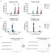
Construction of recombinant VSV with amino acid mutations in M protein. (A,B) Schematic representation of M protein mutant rVSVs construction. Plasmids with mutations in the VSV M gene were constructed using reverse genetics. Genomic plasmids (pVSV-FL-WT, pVSV-FL-M33A/M51R, or pVSV-FL-Mmut) along with helper plasmids (pBS-VSV-N, pBS-VSV-P, pBS-VSV-G, and pBS-VSV-L), were co-transfected into 293T cells pre-infected with vTF7-3. Subsequently, the pBS-VSV-G plasmid was transfected into Vero cells to rescue the virus, resulting in the construction of three viruses: rVSV-WT, rVSV-M2, and rVSV-M4. (C) Agarose gel electrophoresis (AGE) and next generation sequencing technology was used to verify the correctness of the viral M gene. (D) SDS-PAGE (left) and Western Blot (right) analyses were identified the integrity of the viral proteins.
Figure 2
Figure 2
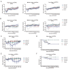
Comparison of the replication capacity of rVSV-WT, rVSV-M2, and rVSV-M4. (A) BHK21 or A549 cells were infected with rVSV-WT, rVSV-M2, or rVSV-M4 separately (MOI of 0.0001). Virus supernatants were harvested at 12, 24, 48, and 72 h post infection. Viral titers (PFU/mL) were determined by plaque assays. GraphPad Prism was used to analyze the significant differences using the one-way ANOVA method. ****, p < 0.0001; ns, p > 0.05. (B) Plaque assays were conducted to compare the replication capabilities of the three viruses in A549, Vero, and Huh7 cells. (C) Ten independent plaques formed by each virus were randomly selected, and plaque areas were quantified using ImageJ 1.8.0 software (National Institutes of Health, Bethesda, MD, USA). GraphPad Prism was used to analyze the significant differences in plaque size using the one-way ANOVA method. Data are presented as mean ± standard deviation (SD). ****, p < 0.0001; ns, p > 0.05.
Figure 3
Figure 3
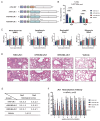
Type I IFN expression enhancement by M protein mutant. (A) Huh-7 and A549 cells were infected with rVSV-WT, rVSV-M2, or rVSV-M4 at MOI of 0.0001. The supernatants were harvested at 12, 24, 48, and 72 h post-infection to detect the levels of INF-α and INF-β. In A549 cells, the Mmut group exhibited the strongest interferon response, whereas the WT group showed the weakest. In Huh-7 cells, INF-α and INF-β levels in all groups were below the detection limit (Figure S1). n = 3; *, p < 0.05; **, p < 0.01; ***, p < 0.001; ****, p < 0.0001. (B) C57BL/6 mice were immunized intranasally with two different doses of rVSV-WT, rVSV-M2, or rVSV-M4 at a single dose of 1 × 10⁷ PFU (left) or 1 × 10⁵ PFU (right). INF-β levels in serum were detected at 12, 24, 48, and 72 h post-infection. n = 6; *, p < 0.05; **, p < 0.01; ****, p < 0.0001. (C) C57BL/6 mice were immunized intranasally at a single dose of 1 × 10⁵ PFU. Mice were sacrificed at various time points., and whole lung tissues were collected for transcriptome sequencing. Differentially expressed genes were enriched and subjected to functional analysis.
Figure 4
Figure 4
Comparison of the safety evaluation via intranasal and intracranial inoculation. (AF): Female C57BL/6 mice aged 6–8 weeks were selected for immunization experiments. n = 6. Data are presented as mean ± standard deviation (SD). (A) Primary immunization: C57BL/6 mice were intranasally inoculated with a high (1 × 10⁷ PFU), medium (1 × 10⁵ PFU), or low (1 × 10³ PFU) dose of rVSV-WT, rVSV-M2, or rVSV-M4, respectively. The vehicle group received PBS, while the MOCK group remained untreated. Body weight changes were monitored over a 21-day period. Mice infected with rVSV-M2 or rVSV-M4 exhibited less weight loss compared to those infected with rVSV-WT. Less weight loss for M2 and M4 viruses only occurred at the 1 × 10³ and 1 × 10⁵ pfu experiments because at 1 × 10⁷, all VSV vaccinated mice lost weight early after vaccination. (B) Challenge post-immunization: Immunized C57BL/6 mice were challenged with a dose of 1 × 10⁷ PFU of rVSV-WT, while the MOCK group remained untreated. Body weight changes were recorded over 21 days. The protective efficacy of the M protein mutant rVSVs was comparable to that of rVSV-WT in C57BL/6 mice. (C–F): C57BL/6 mice were intracranially inoculated with high (1 × 10⁷ PFU) or low (1 × 10³ PFU) dose of rVSV-WT, rVSV-M2, or rVSV-M4, respectively, while the vehicle group received PBS. Body weight changes and survival rates were monitored over a 21-day period.
Figure 5
Figure 5
Comparison of inflammatory responses induced by rVSV-WT, rVSV-M2, or rVSV-M4 in vivo. *, p < 0.05; **, p < 0.01. (A,B): Female C57BL/6 mice aged 6–8 weeks were selected for immunization experiments. C57BL/6 mice were intranasally inoculated with a single dose of 1 × 10⁷ PFU of rVSV-WT, rVSV-M2, or rVSV-M4, respectively, while the vehicle group received PBS. Blood and serum were collected at various time points. n = 4. Data are presented as mean ± standard deviation (SD). (A) TNF-alpha, IL-6, IFN-gamma, and CCL4 levels in serum were detected at 4, 24, and 48 h post-infection. (B) Complete blood cell counts were performed to determine the numbers of white blood cells (WBC), red blood cells (RBC), and platelets (PLT). Additionally, white blood cells (WBC) were further classified into subpopulations.
Figure 6
Figure 6
Construction of mucosal COVID-19 vaccine candidates based on rVSV. (A) Schematic diagram of viral construction: The VSV G protein was replaced with the COVID-19 spike protein through viral packaging technology, followed by point mutations in the VSV M protein. (BE): Hamsters were intranasally immunized with a single dose of 1 × 10⁵ PFU, while the vehicle group received PBS. Blood and serum were collected at various time points, and hamsters were sacrificed on the second and fifth days post-immunization. n = 4. (B) The viral RNA titers in hamster lung tissue were quantified by RT-qPCR. (C) Complete blood cell counts were performed to determine and classify white blood cells. (D) Lung tissues in hamsters on day 2 and 5 post-infection were subjected to H&E staining to observe pathological changes. (E) Histological scoring of hamster lung tissue was conducted as follows: 0: Intact alveolar walls without thickening, no inflammatory infiltration, no congestion. 1: Mild diffuse inflammatory cell (neutrophil) infiltration in alveolar walls, no significant thickening. 2: Prominent and widespread inflammatory cell infiltration (neutrophils and monocytes), mild thickening of alveolar walls (1–2 times). 3: Severe inflammatory cell infiltration, localized thickening of alveolar walls (3–5 times). 4: Severe inflammatory cell infiltration, significant thickening of alveolar walls, 25–50% of lung tissue consolidated. 5: Severe inflammatory cell infiltration, significant thickening of alveolar walls, >50% of lung tissue consolidated. (F) Hamsters were intranasally immunized with two doses (1 × 10⁶ PFU per dose) of rVSV-JN.1, rVSV-M2-JN.1, or rVSV-M4-JN.1 on day0 and day21, while the vehicle group received PBS. Neutralizing antibody titers in serum were detected using a previously established pseudovirus neutralization platform. Nonlinear fitting was performed using GraphPad Prism 9.4.1 to calculate the ID50.

Similar articles

References

    1. Kumar S.U., Priya N.M., Nithya S.R., Kannan P., Jain N., Kumar D.T., Magesh R., Younes S., Zayed H., Doss C.G.P. A review of novel coronavirus disease (COVID-19): Based on genomic structure, phylogeny, current shreds of evidence, candidate vaccines, and drug repurposing. 3 Biotech. 2021;11:198. doi: 10.1007/s13205-021-02749-0. - DOI - PMC - PubMed
    1. Iwasaki A., Omer S.B. Why and How Vaccines Work. Cell. 2020;183:290–295. doi: 10.1016/j.cell.2020.09.040. - DOI - PMC - PubMed
    1. Lin K., Zhong X., Ying M., Li L., Tao S., Zhu X., He X., Xu F. A mutant vesicular stomatitis virus with reduced cytotoxicity and enhanced anterograde trans-synaptic efficiency. Mol. Brain. 2020;13:45. doi: 10.1186/s13041-020-00588-3. - DOI - PMC - PubMed
    1. Lan Q., Wang L., Jiao F., Lu L., Xia S., Jiang S. Pan-coronavirus fusion inhibitors to combat COVID-19 and other emerging coronavirus infectious diseases. J. Med. Virol. 2022;95:e28143. doi: 10.1002/jmv.28143. - DOI - PMC - PubMed
    1. Fuchs J.D., Frank I., Elizaga M.L., Allen M., Frahm N., Kochar N., Li S., Edupuganti S., Kalams S.A., Tomaras G.D., et al. First-in-Human Evaluation of the Safety and Immunogenicity of a Recombinant Vesicular Stomatitis Virus Human Immunodeficiency Virus-1 gag Vaccine (HVTN 090) Open Forum Infect. Dis. 2015;2:ofv082. doi: 10.1093/ofid/ofv082. - DOI - PMC - PubMed

MeSH terms

LinkOut - more resources