. 2025 Aug 14;17(8):1120.
doi: 10.3390/v17081120.

Identification and Structural Characterization of Viroporins from Deadly Hemorrhagic Viruses

Affiliations

Identification and Structural Characterization of Viroporins from Deadly Hemorrhagic Viruses

Hiya Lahiri et al. Viruses. .

Abstract

Crimean-Congo hemorrhagic fever virus (CCHF-V) and Ebola virus are lethal pathogens that cause widespread outbreaks of hemorrhagic fever. Both diseases can be transmitted through contact with the bodily fluids of infected individuals, but as an arbovirus, CCHF-V is primarily transmitted through tick bites. Both of these viruses are classified as Risk Group 4 due to the appreciable health threat they pose. To date, there are few effective treatments available to combat these deadly hemorrhagic fevers. Consequently, identifying and characterizing ion channels (viroporins) encoded in the viral genomes may lead to potential targeted drug development. Therefore, using bacteria-based genetic assays, two viroporin candidates from CCHF-V and Ebola have been examined, and their proposed structures have been modeled to aid in further drug discovery. The results indicate that CCHF-V-gp exhibits channel activity, which is indistinguishable from established viroporins found in other viruses. In contrast, our experimental approach was unable to uncover a viroporin candidate in the Ebola virus.

Keywords: Crimean–Congo hemorrhagic fever; Ebola virus disease; ion channels; structural analysis; viroporins.


Conflict of interest statement

The authors declare no conflicts of interest.

Figures
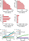
Figure 1
Figure 1
Three bacteria-based assays to asses the channel activity of CCHF-V-gp protein (ac) and Ebola virus delta peptide (df): negative assay panels (a,d); positive assay panels (b,e); fluorescence-pH assay panels (c,f).
Figure 2
Figure 2
Modeling of CCHF-V-gp. (a) lDDT score per residue for predicted oligomers obtained from AlphaFold2-Colab [46]. Five different structural models of different ranks are presented for each oligomer. (b) Side and top views of the highest ranking structure of each oligomer.
Figure 3
Figure 3
Structure of the pores and continuous channels present in the CCHF-V-gp tetramer (a) and pentamer (b). The tetramer shows only discrete pore topology, whereas the pentamer has a continuous channel geometry (shown in the left hand side of each structure).
Figure 4
Figure 4
(a) MD–simulated structure of the CCHF-V-gp depicting charged residues in blue and hydrophobic residues in yellow. (b) Ramachandran plot of amino acid residues indicating the different geometries present in the structure. Each point was obtained by averaging the angles obtained from the molecular dynamics trajectory from 20 to 100 ns. (c) RMSD versus time plot of protein backbone atoms over 100 ns trajectory. (d) RMSF per residue plot for the protein obtained from the last 50 ns of the trajectory. The transmembrane (TM) domain has been marked within the curve. The green shaded region signifies the helical geometry.
Figure 5
Figure 5
Workflow showing the overall process of identifying and characterizing viroporins employed in the current studies.
Figure 6
Figure 6
(a) Pro12 residue within the transmembrane domain forms a kink in helical bundles to widen the channel structure. (b) Transmembrane domain helical bundles from the cytosolic view point, depicting the outwards orientation of Arg24 sidechain.
Figure 7
Figure 7
(a) β–turn of the Ebola delta peptide C-terminal showing the vicinity of the two Cys residues. (b) Ramachandran plot of the β-turn zone showing the dihedral angles. (c) Sequence alignment of different Ebola viruses showing the conserved residues in color. The two S-S bond-forming Cys residues are marked in yellow. * (asterisk): All sequences have the exact same amino acid at this position (fully conserved). : (colon): All amino acids at this position have highly similar properties; . (period): Amino acids have moderately similar properties.

Similar articles

References

    1. Fields B.N. Fields’ Virology. Volume 1 Lippincott Williams & Wilkins; Philadelphia, PA, USA: 2007.
    1. Hoogstraal H. The epidemiology of tick-borne Crimean-Congo hemorrhagic fever in Asia, Europe, and Africa. J. Med. Entomol. 1979;15:307–417. doi: 10.1093/jmedent/15.4.307. - DOI - PubMed
    1. Ergonul O., Whitehouse C.A. Crimean-Congo Hemorrhagic Fever: A Global Perspective. Springer Science & Business Media; Berlin/Heidelberg, Germany: 2007.
    1. Blair P.W., Kuhn J.H., Pecor D.B., Apanaskevich D.A., Kortepeter M.G., Cardile A.P., Ramos A.P., Keshtkar-Jahromi M. An emerging biothreat: Crimean-Congo hemorrhagic fever virus in southern and western Asia. Am. J. Trop. Med. Hyg. 2019;100:16. doi: 10.4269/ajtmh.18-0553. - DOI - PMC - PubMed
    1. Garrison A.R., Alkhovsky S.V., Avšič-Županc T., Bente D.A., Bergeron É., Burt F., Di Paola N., Ergünay K., Hewson R., Kuhn J.H., et al. ICTV virus taxonomy profile: Nairoviridae. J. Gen. Virol. 2020;101:798–799. doi: 10.1099/jgv.0.001485. - DOI - PMC - PubMed

MeSH terms

Substances

LinkOut - more resources