. 2025 Jul 23;13(8):780.
doi: 10.3390/vaccines13080780.

Systemic and Mucosal Humoral Immune Responses to Lumazine Synthase 60-mer Nanoparticle SARS-CoV-2 Vaccines

Affiliations

Systemic and Mucosal Humoral Immune Responses to Lumazine Synthase 60-mer Nanoparticle SARS-CoV-2 Vaccines

Cheng Cheng et al. Vaccines (Basel). .

Abstract

Background: Vaccines that stimulate systemic and mucosal immunity to a level required to prevent SARS-CoV-2 infection and transmission are an unmet need. Highly protective hepatitis B and human papillomavirus nanoparticle vaccines highlight the potential of multivalent nanoparticle vaccine platforms to provide enhanced immunity. Here, we report the construction and characterization of self-assembling 60-subunit icosahedral nanoparticle SARS-CoV-2 vaccines using the bacterial enzyme lumazine synthase (LuS). Methods and Results: Nanoparticles displaying prefusion-stabilized SARS-CoV-2 spike ectodomains fused to the surface-exposed amino terminus of LuS were designed using structure-guided approaches. Negative stain-electron microscopy studies of purified nanoparticles were consistent with self assembly into 60-mer nanoparticles displaying 20 spike trimers. After two intramuscular doses, these purified spike-LuS nanoparticles elicited significantly higher SARS-CoV-2 neutralizing activity than spike trimers in vaccinated mice. Furthermore, intramuscular DNA priming and intranasal boosting with a SARS-CoV-2 LuS nanoparticle vaccine stimulated mucosal IgA responses. Conclusion: These data identify LuS nanoparticles as highly immunogenic SARS-CoV-2 vaccine candidates and support the further development of this platform against SARS-CoV-2 and its emerging variants.

Keywords: SARS-CoV-2; lumazine synthase; mucosal immunity; nanoparticle; vaccine.


Conflict of interest statement

The authors declare no conflicts of interest.

Figures
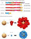
Figure 1
Figure 1
Design and molecular modeling of vaccine constructs. (A) Schematic representation of the S-2P full-length trimer. A “GSAS” substitution (yellow box) eliminates cleavage by furin between S1 and S2 domains (residues 682–685). Proline substitutions at residues 986 and 987 stabilize the S protein in the prefusion conformation. Amino acid (AA) positions for known S subunits are labelled on the top of the construct: amino acid 1-19: signal peptide; NTD: N-terminal domain; RBD: receptor binding domain; SD1/2: subdomains 1 and 2; S2: S2 domain; The full length of S also include TM: transmembrane domain; Cyto: cytoplasmic domain. The S-2P constructs contain two proline mutations at amino acid positions 986 and 987, while the S-6P constructs contain four additional proline mutations (F817P, A892P, A899P, A942P), as indicated by the purple boxes. The ecotodomain of S from amino acid 1-1147 or 1-1206 is fused to the N-terminus of lumazine synthase (LuS) in S-2P-1147-LuS or S-2P-1206-LuS, respectively. NTD-LuS contains the NTD and LuS fusion. (B) Molecular modeling of S-2P-LuS and NTD-LuS nanoparticles. Constructs were modeled using existing structures of the S-2P trimer (PDB ID 6VXX) and LuS 60-mer (PDB ID 1HQK). 3AA linker: GSG; 7AA linker: GSGGGSG; 15AA linker: GGSGGSGGSGGSGGG; Linker for NTD-LuS: GGSGGSGGSGGSGG.
Figure 2
Figure 2
Antigenic characterization of nanoparticles. (A) Four RBD-specific antibodies (2-4, LY-CoV555, S309, and H4), one NTD antibody (4-8), and ACE2 fused with hFc were used to assess the antigenicity of these nanoparticles. S-2P trimers, RBD monomers, and NTD monomer proteins were included in these studies as control proteins. (B) Antigenic characterization of SARS-CoV-2 Beta variant B.1.351 and S-6P-LuS14 using two RBD mAbs (LY-CoV555, S-309), one NTD mAb (4-8), and ACE2 fused with hFc.
Figure 3
Figure 3
Immunogenicity of SARS-CoV-2 LuS Nanoparticle Immunogens in Mice. (A) Immunization scheme for groups of mice (n = 5 per group) immunized with different doses (0.08, 0.4 or 2 ug) of nanoparticle or recombinant trimer immunogens. (B) The S-2P protein binding or (C) RBD-binding IgG response as measured by ELISA as shown at Week 2 (left panel) and 5 (right panel). (D) Serum neutralization of a SARS-CoV-2 Wuhan strain pseudotype was measured with geometric means for neutralization titers shown for each group. Data (BD) were analyzed by Kruskal–Wallis test followed by Dunn’s multiple comparison test. (* p  <  0.05; ** p  <  0.01). Dashed lines represent the lowest serum dilution at 1:40 in the neutralization assay as the detection limits.
Figure 4
Figure 4
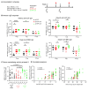
Potent immune responses against Wuhan WT and VOC were elicited in mice. (A) Immunization scheme for four groups of mice (n = 5) with 1ug of the indicated immunogens. (B) The elicited IgG response as measured by ELISA is shown at Week 5 (post 2nd immunization) to SARS-CoV-2 spike (left panels), RBD (middle panel), and NTD (right panel) antigens. (C) Week 5 serum neutralization activity (ID50) against Wuhan WT SARS-CoV-2 pseudovirus. (D) Serum anti-S-2P spike IgG titers time course from Weeks 0 to 40. (E) serum anti-SARS-CoV 2 S-2P IgG responses pre and post S-6P LuS B.1.351 boost. (F) Serum neutralization activity (ID50) against pseudotyped Wuhan WT SARS-CoV-2 and VoC (alpha, beta, delta) at pre (wk31) and post (wk 40) S-6P Beta Variant boost. Geometric mean and 95% CI were shown. Data for panel (B) were analyzed by Kruskal–Wallis test followed by Dunn’s multiple comparison test, data (E,F) were analyzed with two-tailed Mann–Whitney test. (* p  <  0.05; ** p  <  0.01).
Figure 5
Figure 5
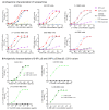
DNA delivery of S nanoparticle immunogens elicited immune responses. (A) Immunization scheme for nine groups of mice (n = 10) immunized with either 10, 2, or 0.4 µg of indicated DNA plasmids twice intramuscularly. (B) SARS-CoV-2-S2P and SARS-CoV-2-RBD IgG response assessed by ELISA at Week 2 or Week 5. (C) Week 5 serum neutralization and (D) two-sided Pearson correlation of anti-RBD (left panel) or anti-S-2P (right panel) IgG ELISA endpoint titer to SARS-CoV-2 Wuhan 1 WT neutralization titer. Data (B,C) were analyzed by Kruskal–Wallis test followed by Dunn’s multiple comparison test. (* p  <  0.05; ** p  <  0.01; *** p  <  0.001; **** p  <  0.0001).
Figure 6
Figure 6
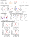
IM DNA Immunization and protein IN boost elicit systemic and mucosal immune response. (A) Extended schedule for the study shown in Figure 5. The 10 µg DNA and the 2 µg DNA groups were further boosted at Week 36 with a third IM DNA or 2 μg IN protein boost, respectively. (B) The serum-neutralizing activity against pseudotyped SARS-CoV-2 Wuhan virus at Week 38 is shown. (C) Mucosal IgG and IgA response at Week 38 as measured with ELISA. IgG response and IgA response were measured at 1:3 dilution of nasal wash or at 1:6 dilution of BAL. Geometric mean and 95% CI are shown for neutralizing activity, mean and SEM are shown in C. Data were analyzed by Kruskal–Wallis test followed by Dunn’s multiple comparison test. (* p  <  0.05; ** p  <  0.01).

References

    1. World Health Organization WHO Coronavirus (COVID-19) Dashboard. [(accessed on 7 February 2025)]. Available online: https://data.who.int/dashboards/covid19/
    1. Barouch D.H. Covid-19 Vaccines—Immunity, Variants, Boosters. N. Engl. J. Med. 2022;387:1011–1020. doi: 10.1056/NEJMra2206573. - DOI - PMC - PubMed
    1. Walls A.C., Park Y.J., Tortorici M.A., Wall A., McGuire A.T., Veesler D. Structure, Function, and Antigenicity of the SARS-CoV-2 Spike Glycoprotein. Cell. 2020;181:281–292.e6. doi: 10.1016/j.cell.2020.02.058. - DOI - PMC - PubMed
    1. Wrapp D., Wang N., Corbett K.S., Goldsmith J.A., Hsieh C.L., Abiona O., Graham B.S., McLellan J.S. Cryo-EM structure of the 2019-nCoV spike in the prefusion conformation. Science. 2020;367:1260–1263. doi: 10.1126/science.abb2507. - DOI - PMC - PubMed
    1. Cai Y., Zhang J., Xiao T., Peng H., Sterling S.M., Walsh R.M., Jr., Rawson S., Rits-Volloch S., Chen B. Distinct conformational states of SARS-CoV-2 spike protein. Science. 2020;369:1586–1592. doi: 10.1126/science.abd4251. - DOI - PMC - PubMed

LinkOut - more resources