. 2025 Jul 28;13(8):802.
doi: 10.3390/vaccines13080802.

Development and Protective Efficacy of a Novel Nanoparticle Vaccine for Gammacoronavirus Avain Infectious Bronchitis Virus

Affiliations

Development and Protective Efficacy of a Novel Nanoparticle Vaccine for Gammacoronavirus Avain Infectious Bronchitis Virus

Ting Xiong et al. Vaccines (Basel). .

Abstract

Background: Infectious bronchitis virus (IBV) is a gammacoronavirus that causes a highly contagious disease in chickens and seriously endangers the poultry industry. The GI-19 is a predominant lineage. However, no effective commercially available vaccines against this virus are available. Methods: In this present study, the CHO eukaryotic and the E.coli prokaryotic expression system were used to express S1-SpyTag and AP205-SpyCatcher, respectively. Subsequently, the purified S1-SpyTag and AP205-SpyCatcher were coupled to form the nanoparticles AP205-S1 (nAP205-S1) in PBS buffer at 4 °C for 48 h. S1-SpyTag and nAP205-S1 were formulated into vaccines with white oil adjuvant and employed to immunize 1-day-old SPF chickens for the comparative evaluation of their immune efficacy. Results: The nAP205-S1 vaccine in chickens induced robust IBV-specific humoral and cellular immune responses in vivo. Importantly, the humoral and cellular immune responses elicited by the nAP205-S1 vaccine were more robust than those induced by the IBV S1-SpyTag vaccine at both the same dose and double the dose, with a notably significant difference observed in the cellular immune response. Furthermore, experimental data revealed that chicken flocks vaccinated with nAP205-S1 achieved 100% group protection following a challenge, exhibiting a potent protective immune response and effectively inhibiting viral shedding. Conclusions: These results reveal the potential of developing a novel nanoparticle vaccine with broadly protective immunity against GI-19 IBV.

Keywords: IBV; nanoparticle vaccine; protective efficacy.


Conflict of interest statement

Authors Yanfen Lyu and Ruiai Chen were employed by the company Zhaoqing Dahuanong Biology Medicine Co., Ltd. The remaining authors declare that the research was conducted in the absence of any commercial or financial relationships that could be construed as a potential conflict of interest.

Figures
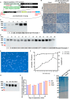
Figure 1
Figure 1
The IBV S1-SpyTag protein was synthesized through serum-free suspension culture technology of CHO cells. (A) Schematic diagram of the pXJ40 (NeoR/KanR)+-S1-SpyTag expression plasmid. (B) The G418 drug screening approach for positive cells. The cell states of the recombinant plasmid-transfected group and empty vector-transfected group at the 1st, 4th, and 8th days post-transfection. (C) Western blot analysis was used to compare the protein expression levels in the supernatants of different CHO-S1 cell clusters during expansion culture using monoclonal antibody anti-IBV S and goat anti-mouse IgG-HRP. (D) Western blot analysis was used to compare the protein expression levels in the supernatants of different monoclonal CHO-S1 cell lines during expansion culture using monoclonal antibody anti-IBV S and goat anti-mouse IgG-HRP. (E) Diagram illustrating the cell state of CHO stable cell line during serum-free suspension adaptation. (F) Monitoring of growth density and viability of serum-free fully suspended CHO stable cells. (G) The expression stability of the CHO stable cell line expressing IBV S1-SpyTag was evaluated. Western blot analysis was performed to detect the levels of secreted protein in the supernatant of the CHO stable cell line at passages 1, 5, 10, and 15 during suspension culture, corresponding to p1, p5, p10, and p15, respectively. (H) The differences in the expression levels of IBV S1-SpyTag in CHO stable cell lines were compared among three different serum-free media: 031, Han1, and Han2. (I) SDS-PAGE analysis of the purity of IBV S1-SpyTag purified protein using 10% gel.
Figure 2
Figure 2
The AP205 coat protein was efficiently synthesized in E. coli. (A) Schematic diagram of the pET 28(b)+-AP205-SpyCathcer expression plasmid. (B) SDS-PAGE analysis of the expression level of AP205-SpyCatcher protein in supernatant and precipitation using 10% gel. The AP205-SpyCatcher construct was transformed into E. coli BL21(DE3), which was first grown at 37 °C in LB broth containing kanamycin to OD600 = 0.6~0.8 and then induced with 0.5 mM IPTG for 19 h at 16 °C. (C) SDS-PAGE analysis of the purity of IBV S1-SpyTag purified protein using 10% gel. The supernatant containing the synthesized AP205-SpyCatcher protein was filtered through a 0.45 μm filter membrane. The filtrate was then loaded onto a Ni-NTA affinity column and incubated overnight at 4 °C with gentle inversion to ensure efficient binding of the target protein.
Figure 3
Figure 3
Generation of nAP205-S1 and vaccine preparations. (A) Schematic illustration of the in vitro chemical conjugation and assembly of nAP205-S1 nanoparticles mediated by AP205-SpyCatcher and IBV S1-SpyTag. (B) SDS-PAGE analysis of the in vitro assembly efficiency of AP205-S1 nanoparticles using 10% gel. nAP205-S1 is a nanoparticle antigen assembled from AP205-SpyCatcher and IBV S1-SpyTag. (C) The shape of nAP205-S1 nanoparticles was detected and analyzed by electron microscopy. (D) Three vaccines solutions were formulated containing conjugated nAP205-S1 or IBV S1-SpyTag. nAP205-S1 or IBV S1-SpyTag vaccines were at concentrations of 6 μg/0.2 mL, 6 μg/0.2 mL, and 12 μg/0.2 mL, respectively.
Figure 4
Figure 4
Humoral and cellular immune efficiencies in chickens immunized with nAP205-S1. (A) Immunization and exsanguination protocol. Prime-immunization with PBS, IBV S1-SpyTag (6 µg/0.2 mL), IBV S1-SpyTag (12 μg/0.2 mL), and nAP205-S1 (6 μg/0.2 mL) vaccines, respectively. Boost-immunization at 10 days, respectively. Blood samples were collected weekly after immunization. (B) Serum antibody titers against infectious bronchitis virus (IBV) were assessed using ELISA at the specified time points. (C) The proportions of CD3+, CD4+, and CD8+ T cell populations in peripheral blood samples collected at 28 days post-infection were analyzed by flow cytometry, with statistical evaluation performed using GraphPad Prism 9. The levels of statistical significance are indicated by p values, where ns represents non-significance, * p < 0.05, *** p < 0.001 and **** p < 0.0001.
Figure 5
Figure 5
Immune protection efficiency against CK/CH/JS/TAHY. (A) Survival curves of six representative viral isolates were generated using 1-day-old SPF chickens. Each group of birds was inoculated via the nasal–ocular route with approximately ~105.5 EID50 of one of the six selected isolates. The number of mortalities occurring within 8 days post-challenge was recorded and survival curves were plotted using the Graphpad Prism 9 program. The survival curves of IBV S1-SpyTag (6 µg/0.2 mL), IBV S1-SpyTag (12 µg/0.2 mL), nAP205-S1 (6 µg/0.2 mL), and PBS Con are represented by pink, orange, blue, and black lines, respectively. (B) At 8 days post-challenge with CK/CH/JS/TAHY, trachea, lung, and kidney autopsies of dead chickens and all surviving chickens from each experimental group. Red and blue arrows indicate the pathological lesions observed. (C) At 8 days post-challenge with CK/CH/JS/TAHY, pathological lesions in the trachea, lung, and kidney autopsies of unvaccinated and unchallenged chickens in the same isolators.
Figure 6
Figure 6
Histopathological analysis of the trachea, lungs, and kidneys was performed on chickens challenged with CK/CH/JS/TAHY. (A) Microscopic examination was carried out on tracheal, pulmonary, and renal tissues obtained from deceased chickens or all surviving birds in each experimental group at 8 days post-challenge. (B) The pathological tissue change scores of the trachea, lungs, and kidneys in each immune-challenged group were statistically analyzed using Graphpad Prism 9 program at 8 days post-challenge. The levels of statistical significance are indicated by p values, where ns represents non-significance and * p < 0.05.

Similar articles

References

    1. Wang Y., Grunewald M., Perlman S. Coronaviruses: An Updated Overview of Their Replication and Pathogenesis. Methods Mol. Biol. 2020;2203:1–29. doi: 10.1007/978-1-0716-0900-2_1. - DOI - PMC - PubMed
    1. Fung T.S., Liu D.X. Human Coronavirus: Host-Pathogen Interaction. Annu. Rev. Microbiol. 2019;73:529–557. doi: 10.1146/annurev-micro-020518-115759. - DOI - PubMed
    1. Legnardi M., Tucciarone C.M., Franzo G., Cecchinato M. Infectious Bronchitis Virus Evolution, Diagnosis and Control. Vet. Sci. 2020;7:79. doi: 10.3390/vetsci7020079. - DOI - PMC - PubMed
    1. Jackwood M.W. Review of infectious bronchitis virus around the world. Avian Dis. 2012;56:634–641. doi: 10.1637/10227-043012-Review.1. - DOI - PubMed
    1. Xiong T., Xie H., Li L., Liang S., Huang M., Yu C., Zhuang T., Liang X., Liu D., Chen R. Prevalence, Genotype Diversity, and Distinct Pathogenicity of 205 Gammacoronavirus Infectious Bronchitis Virus Isolates in China during 2019–2023. Viruses. 2024;16:930. doi: 10.3390/v16060930. - DOI - PMC - PubMed

LinkOut - more resources