. 2025 Sep;77(5):e70072.
doi: 10.1111/jpi.70072.

Melatonin Induces PERK-ATF4 Unfolded Protein Response and Apoptosis in Human Choriocarcinoma Cells

Affiliations

Melatonin Induces PERK-ATF4 Unfolded Protein Response and Apoptosis in Human Choriocarcinoma Cells

Josianne Bienvenue-Pariseault et al. J Pineal Res. 2025 Sep.

Abstract

Melatonin, an indolamine primarily recognized for regulating circadian rhythms, has also demonstrated notable antitumoral properties. Melatonin induces endoplasmic reticulum (ER) stress, modulates autophagy, and promotes apoptosis in various tumors, including gastric, ovarian, cervical, oral tongue, colorectal, renal, hepatic, and bladder cancer. In placental choriocarcinoma, melatonin reduces cell viability and induces apoptosis by inhibiting autophagy and disrupting the mitochondrial membrane potential. However, its effects on ER stress and the unfolded protein response (UPR) pathway remain unexplored. It is hypothesized here that the proapoptotic effects of melatonin in choriocarcinoma cells occur through the activation of the UPR pathway. The factors implicated in the UPR (PERK, IRE1ɑ, ATF6, GRP78, ATF4, CHOP, P-eIF2α) pathways were evaluated by Western blot, RT-qPCR, and flow cytometry in BeWo (human choriocarcinoma) cells treated with or without melatonin (1 mM). Melatonin significantly increased protein levels of GRP78 (p = 0.0329), IRE1α (p = 0.0394), p-eIF2α (p = 0.0439), ATF4 (p = 0.0267), CHOP (p = 0.0379), Bax and cleaved PARP but did not affect TRAF2 and NFkB protein levels nor XBP1 mRNA splicing. PERK knockdown, via siRNA, prevented the rise in GRP78, p-eIF2α/eIF2α, and ATF4 levels by melatonin. Additionally, melatonin increased early apoptosis in BeWo cells (p = 0.0371) and PERK knockdown increased the susceptibility of BeWo cells to apoptosis when treated with tunicamycin (p = 0.0359), suggesting that ER stress plays a role in BeWo cell survival. This study demonstrates that melatonin activates the PERK-ATF4-P-eIF2α-CHOP pathway and induces early apoptosis in BeWo cells, while PERK deficiency compromises cell survival under ER stress. Our findings suggest that modulating PERK-UPR signaling with melatonin could present a promising therapeutic strategy for cancer, including placental choriocarcinoma.

Keywords: BeWo cell; PERK pathway; apoptosis; choriocarcinoma; indolamine; melatonin; unfolded protein response.


Conflict of interest statement

The authors declare no conflicts of interest.

Figures

Figure 1
Figure 1
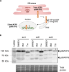
Melatonin increases unfolded protein (UPR) response in BeWo cells. BeWo cells were treated with the vehicle control (0.1% DMSO) with or without melatonin (1 mM) or Tm (3.5 µg/mL) under normoxia (8% O2) conditions for 24‐h. GRP78 protein level was determined by Western blot. Equal protein amounts of cell lysates (35 µg) were subjected to Western blot assay using anti‐GRP78. Total protein was used for normalization. GRP78: 78‐kDa glucose‐regulated protein; DMSO: dimethylsulfoxyde; Mel: melatonin; Tm: municamycin; Data are shown as mean ± SD and were analyzed using an unpaired t‐test (DMSO vs Mel, *p < 0.05), n = 5. To facilitate readers' experiences, the certain band order has been changed. Those modifications have been identified with dotted lines. Original western blots are included under Figure S2.
Figure 2
Figure 2
Melatonin has no effect on the protein level of factors implicated in IREα‐UPR pathway. BeWo cells were treated with the vehicle (DMSO 0.1%) or melatonin (1 mM) or Tm (3.5 μg/mL) under normoxia (8% O2) conditions during 24‐h. (A) IREα, (B) TRAF2 and (C) IkB/NFkB ratio protein level was determined by Western blot. Equal protein amounts of cell lysates (35 µg) were subjected to Western blot assay using anti‐IREα, anti‐TRAF2, anti‐NFkB and anti‐IkB. Total protein was used for normalization. (D) XBP1s/XBP1 gene expression was determined by RT‐qPCR using specific primers. B2M and SDHA were used as references genes for normalization. DMSO: dimethylsulfoxyde; IkB: inhibitor of nuclear factor kappa B; IREα: serine/threonine‐protein kinase/endoribonuclease inositol‐requiring enzyme 1α; Mel: melatonin; NFkB: nuclear factor‐kappa B; TRAF2: TNF receptor‐associated factor 2; Tm: Tunicamycin; XBP1: X‐box binding protein 1; XBP1s: spliced X‐box binding protein 1; Data are shown as mean ± SD and were analyzed using an unpaired t‐test (DMSO vs Mel, *p < 0.05), n = 4–5. To facilitate readers' experiences, the band order of certain blots has been changed. Those modifications have been identified with dotted lines. Original western blots are included under Figures S5–S8.
Figure 3
Figure 3
Melatonin has no effect on ATF6 protein cleavage. BeWo cells were treated with the vehicle (DMSO 0.1%) or melatonin (1 mM) or Tm (3.5 μg/mL) under normoxia (8% O2) conditions during 24‐h. (A) Following GRP78 dissociation, ATF6 (p90ATF6, total ATF6) translocates to the Golgi apparatus, where it undergoes proteolytic cleavage by site‐1 protease (S1P or MBTPS1) and site‐2 protease (S2P or MBTPS2). The resulting activated form, p50ATF6, then enters the nucleus to promote the transcription of genes containing ER stress‐response elements (ERSE‐1). (B) ATF6 protein cleavage level was determined by Western blot. Equal protein amounts of cell lysates were subjected to Western blot assay using anti‐ATF6 antibody. Total protein was used for normalization. ATF6, activating transcription factor 6; DMSO, dimethylsulfoxyde; ER, endoplasmic reticulum; GRP78, 78‐kDa glucose‐regulated protein; Mel, melatonin; S1P, site‐1 protease; S2P, site‐1 protease; Tm, tunicamycin.
Figure 4
Figure 4
Melatonin increases the protein level of factors implicated in PERK‐UPR pathway. BeWo cells were treated with the vehicle (DMSO 0.1%) or melatonin (1 mM) or Tm (3.5 μg/mL) under normoxia (8% O2) conditions during 24‐h. (A) p‐eif2α, (B) eif2α, (C) ATF4, (D) CHOP, (E) Bax, (F) Bcl‐2, and (G) cl‐Parp protein level was determined by Western blot. Equal protein amounts of cell lysates (35 µg) were subjected to Western blot assay using anti‐p‐eif2α, anti‐eif2α, anti‐ATF4, anti‐CHOP, anti‐Bax, anti‐Bcl‐2 and anti‐cl‐Parp. Total protein was used for normalization. ATF4, activating transcription factor 4; Bax, Bcl‐2–associated X; Bcl‐2, B‐cell lymphoma 2; CHOP, DNA damage‐inducible transcript 3, also known as C/EBP homologous protein; cl‐Parp, cleaved poly (ADP‐ribose) polymerase; DMSO, dimethylsulfoxyde; eif2α, eukaryotic initiation factor 2 subunit alpha; Mel, melatonin; p‐eif2α, phosphorylation of the eukaryotic initiation factor 2 subunit alpha; Tm, tunicamycin. Data are shown as mean ± SD and were analyzed using an unpaired t‐test (DMSO vs. Mel, *p < 0.05), n = 4–6. To facilitate readers' experiences, the band order of certain blots has been changed. Those modifications have been identified with dotted lines. Original western blots are included under Figures S12–S18.
Figure 5
Figure 5
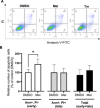
Melatonin increases early apoptosis in BeWo cells. BeWo cells were treated with the vehicle (DMSO) or melatonin (1 mM) or Tm (3.5 μg/mL) under normoxia (8% O2) conditions during 24‐h. (A) Number of apoptotic cells was determined by FACS using Annexin V and PI staining. (B) Number of early or late apoptotic cells are expressed as a percentage of the control DMSO. Ann, annexin V; DMSO, dimethylsulfoxyde; Early, early apoptosis; Late, late apoptosis; Mel, melatonin; PI, propidium iodide; Tm, tunicamycin; Total, total apoptosis. Data are shown as mean ± SD and were analyzed using an unpaired t‐test (DMSO vs. Mel, *p < 0.05), n = 7.
Figure 6
Figure 6
Knockdown of PERK inhibits the ability of melatonin to induce PERK‐UPR pathway. BeWo cells subjected to scramble (SC) or siRNA against PERK for 72‐h were treated with the vehicle (DMSO 0.1%) or melatonin (1 mM) or TM (3.5 μg/mL) under normoxia (8% O2) conditions during 24‐h. (A) PERK, (B) GRP78, (C) p‐eif2α/eif2α ratio (D) p‐eif2α, (E) eif2α, and (F) ATF4 protein level was determined by Western blot. Equal protein amounts of cell lysates (35 µg) were subjected to Western blot assay using anti‐PERK, anti‐GRP78, anti‐p‐eif2α, anti‐eif2α ratio and anti‐ATF4. Total protein was used for normalization. ATF4, activating transcription factor 4; DMSO, dimethylsulfoxyde; eif2α, eukaryotic initiation factor 2 subunit alpha; GRP78, 78‐kDa glucose‐regulated protein; Mel, melatonin; p‐eif2α, phosphorylation of the eukaryotic initiation factor 2 subunit alpha; PERK, protein kinase R (PKR)‐like; Tm, tunicamycin. Data are shown as mean ± SD and were analyzed using ANOVA followed by Tukey post hoc test (*p < 0.05), or an unpaired t‐test (SC Tm vs. si Tm, # p < 0.05, ## p < 0.01), n = 5. To facilitate readers' experiences, the band order has been changed. Those modifications have been identified with dotted lines. Original western blots are included under Figures S22–S26.
Figure 7
Figure 7
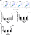
Melatonin and knockdown of PERK have no effect on early and late apoptosis. BeWo cells subjected to scramble (SC) or siRNA against PERK (si) for 72‐h were treated with the vehicle (DMSO 0.1%) or melatonin (1 mM) or Tm (3.5 μg/mL) under 8% of oxygen during 24‐h. (A) Apoptotic cells were determined by FACS using Annexin V and PI. Number of (B) early, (C) late or (D) total apoptotic cells are expressed as a percentage of the control DMSO. Ann, annexin V; DMSO, dimethylsulfoxyde; Early, early apoptosis; Late, late apoptosis; Mel, melatonin; PERK, (PKR)‐like protein kinase R; PI, propidium iodide; Tm, tunicamycin; Total, total (early + late) apoptosis. Data are shown as mean ± SD and were analyzed using ANOVA followed by Tukey post hoc test (*p < 0.05), or an unpaired t‐test (SC Tm vs si Tm, # p < 0.05, ## p < 0.01). n = 5.

Similar articles

References

    1. Schwarz D. S. and Blower M. D., “The Endoplasmic Reticulum: Structure, Function and Response to Cellular Signaling,” Cellular and Molecular Life Sciences 73, no. 1 (2016): 79–94, 10.1007/s00018-015-2052-6. - DOI - PMC - PubMed
    1. Jiang P., Gan M., Lin W.‐L., and Yen S. H., “Nutrient Deprivation Induces α‐Synuclein Aggregation Through Endoplasmic Reticulum Stress Response and SREBP2 Pathway,” Frontiers in Aging Neuroscience 6 (2014): 268, 10.3389/fnagi.2014.00268. - DOI - PMC - PubMed
    1. Sharma V., Kaur R., Bhatnagar A., and Kaur J., “Low‐pH‐Induced Apoptosis: Role of Endoplasmic Reticulum Stress‐Induced Calcium Permeability and Mitochondria‐Dependent Signaling,” Cell Stress and Chaperones 20, no. 3 (2015): 431–440, 10.1007/s12192-014-0568-6. - DOI - PMC - PubMed
    1. Guan L., Ge R., and Ma S., “Newsights of Endoplasmic Reticulum in Hypoxia,” Biomedicine & Pharmacotherapy = Biomedecine & pharmacotherapie 175 (2024): 116812, 10.1016/j.biopha.2024.116812. - DOI - PubMed
    1. Tirasophon W., Welihinda A. A., and Kaufman R. J., “A Stress Response Pathway From the Endoplasmic Reticulum to the Nucleus Requires a Novel Bifunctional Protein Kinase/Endoribonuclease (Ire1p) n Mammalian Cells,” Genes & Development 12, no. 12 (1998): 1812–1824. - PMC - PubMed

MeSH terms

LinkOut - more resources