Review
. 2025 Sep;26(9):841-854.
doi: 10.3348/kjr.2025.0357.

CT and MRI in Advanced Ovarian Cancer: Advances in Imaging Techniques

Affiliations
Review

CT and MRI in Advanced Ovarian Cancer: Advances in Imaging Techniques

Selina Chiu et al. Korean J Radiol. 2025 Sep.

Abstract

Ovarian cancer (OC) remains one of the leading causes of gynecologic cancer-related mortality, with most patients presenting with disseminated disease, particularly within the peritoneal cavity. Standard treatment includes cytoreductive surgery, platinum-based chemotherapy, and targeted maintenance approaches depending on the patient's and tumor's genetic profile. Despite treatment advancements, approximately 25% of high-grade serous OC cases relapse within a year despite optimal primary treatment with complete tumor clearance at cytoreduction. Advances in contrast-enhanced CT (CE-CT) and MRI have revolutionized the evaluation and treatment planning of advanced OC. CT remains the gold standard for staging and assessing tumor extent, effectively identifying peritoneal, lymphatic, and distant metastases. However, it is less effective in detecting small-volume peritoneal dissemination. MRI, with superior soft-tissue contrast, complements CT by providing a detailed assessment of peritoneal disease, characterizing sonographically indeterminate adnexal masses. Diffusion-weighted imaging and gadolinium-enhanced MRI have improved the diagnostic sensitivity for peritoneal disease but are unable to predict treatment response, recurrence risk, and prognosis. Radiomics, which extracts quantitative tumor features from imaging data, holds promise for personalizing treatment and identifying patients at risk for early recurrence despite optimal therapy. The integration of CT, MRI, and radiomics could enhance surgical planning and improve long-term survival outcomes in patients with advanced OC.

Keywords: CT; Lexicon scoring; MRI; Ovarian cancer; Radiomics.


Conflict of interest statement

The authors have no potential conflicts of interest to disclose.

Figures

Fig. 1
Fig. 1. CE-CT and MRI of a patient with high-grade serous ovarian cancer. A, B: CE-CT (A) and DWI-MRI (B) show thickening of the peritoneal reflection in the left paracolic gutter (arrows). C, D: On the wider field of view images from CE-CT (C) and DWI-MRI (D), there are ascites and soft tissue thickening of the right pelvic peritoneal reflection (red arrow, C, D). DWI-MRI shows intense restricted diffusion, in keeping with peritoneal disease (red arrows, D). Note that the intensely restricting adnexal masses are seen in the pelvis (yellow arrows, D). CE = contrast-enhanced, DWI = diffusion-weighted imaging
Fig. 2
Fig. 2. A patient with high-grade serous ovarian cancer and non-specific bowel symptoms following previous primary cytoreductive surgery. A: CE-CT shows thickening of the sigmoid colon (arrow). B, C: The corresponding T2 axial MRI (B) shows isointense soft tissue adherent to the sigmoid colon (arrow), which intensely restricts on DWI-MRI (C) reflecting bowel serosal metastatic deposit (arrow). CE = contrast-enhanced, DWI = diffusion-weighted imaging
Fig. 3
Fig. 3. Large bowel (splenic flexure) disease. Abdominal contrast-enhanced axial CT shows an abnormal soft tissue mass at the splenic flexure with evidence of transmural invasion (arrow). The patient subsequently underwent a de-functioning colostomy.
Fig. 4
Fig. 4. Contrast-enhanced axial CT through the upper abdomen shows a large, necrotic soft tissue mass centered on the gastrohepatic ligament (white arrow) with invasion of the lesser curve of the stomach (yellow arrow) and the liver capsule (red arrow).
Fig. 5
Fig. 5. Typical CE-CT (A) and DWI-MRI (B) appearances of omental deposits, which are intensely restricting in metastatic ovarian cancer (arrows). This is often described as “omental cake.” CE = contrast-enhanced, DWI = diffusion-weighted imaging
Fig. 6
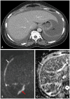
Fig. 6. CE-CT and DWI-MRI of a patient with high-grade serous ovarian cancer. A: The CE-CT shows large volume ascites. Soft tissue along the hepatic capsule is not well appreciated on CE-CT. B, C: DWI-MRI (B) and apparent diffusion coefficient (C) MRI images show restricting soft tissue at the hepatic capsule (white arrows), in keeping with subcapsular disease. Note that there is also restricting peritoneal soft tissue in Morrison's pouch (red arrow). CE = contrast-enhanced, DWI = diffusion-weighted imaging
Fig. 7
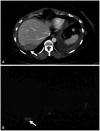
Fig. 7. Liver bare area metastatic disease in the axial plane. A, B: CE-CT (A) and DWI-MRI (B) demonstrate an abnormal soft tissue mass at the liver bare area (arrows). The mass is shown to exhibit diffusion restriction. CE = contrast-enhanced, DWI = diffusion-weighted imaging
Fig. 8
Fig. 8. CE-CT and DWI-MRI of a female patient with metastatic high-grade serous ovarian cancer. A: CE-CT shows ill-defined soft tissue at the porta and along the falciform ligament (arrows) with trace ascites. B: DWI-MRI confirms restricting soft tissue at the porta (red arrow), which encases the portal vein (*). Additionally, there is soft tissue at the falciform ligament (yellow arrow) and in Morrison's pouch (green arrow). The soft tissue at the porta, which encases the portal vein, would render the patient unresectable for primary cytoreductive surgery. CE = contrast-enhanced, DWI = diffusion-weighted imaging
Fig. 9
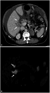
Fig. 9. Porta hepatis in the axial plane. A, B: CE-CT (A) and DWI-MRI (B) through the liver shows abnormal, restricting soft tissue encasing the porta hepatis (arrows). CE = contrast-enhanced, DWI = diffusion-weighted imaging
Fig. 10
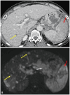
Fig. 10. CE-CT and DWI-MRI of a patient with metastatic high-grade serous ovarian cancer. A: The CE-CT shows soft tissue in the lesser sac (white arrow), which invades the stomach serosa. Hypodense lesions in the liver (yellow arrow) are not fully characterized on CE-CT. The spleen is enlarged and shows heterogeneous enhancement (red arrow). B: DWI-MRI shows extensive restricting soft tissue in the lesser sac (white arrow), more extensive than on CE-CT with invasion into the stomach. Restricting hepatic parenchymal metastases (yellow arrows) are more numerous than on CE-CT. Splenic parenchymal disease (red arrow) is confirmed. CE = contrast-enhanced, DWI = diffusion-weighted imaging
Fig. 11
Fig. 11. CE-CT and DWI-MRI of a patient with metastatic high-grade serous ovarian cancer. A, B: CE-CT (A) and DWI-MRI (B) shows lobulated soft tissue at the splenic hilum (white arrows) and omental nodules just anterior to the spleen (red arrows). DWI-MRI confirms restricting soft tissue at the splenic hilum and omental nodules. Note that on the DWI, restricting subcapsular hepatic disease is well demonstrated (yellow arrow), which was not conspicuous on CE-CT. CE = contrast-enhanced, DWI = diffusion-weighted imaging
Fig. 12
Fig. 12. CE-CT and DWI-MRI images of a patient with metastatic high-grade serous ovarian cancer. A: CE-CT shows a right-sided pleural effusion with the impression of focal pleural enhancement (arrow). B: DWI-MRI images through the thorax confirm restricted diffusion at the right pleura in keeping with pleural metastasis (arrow). CE = contrast-enhanced, DWI = diffusion-weighted imaging
Fig. 13
Fig. 13. CE-CT image of a patient with metastatic high-grade serous ovarian cancer on lung windows shows pulmonary metastases, some of which are cavitating nodules (arrows). CE = contrast-enhanced
Fig. 14
Fig. 14. A summary of LNs in the thorax in patients with metastatic high-grade ovarian cancer on axial CE-CT and DWI-MRI. A-G: LNs at these sites would not be suitable for surgical excision, apart from precardiac nodes in exceptional circumstances. Precardiac LN (A, B), right axillary LNs (C, D), paraesophageal mediastinal LNs (E), paratracheal mediastinal LNs (F), and supraclavicular LNs (G), indicated by arrows. LN = lymph node, CE = contrast-enhanced, DWI = diffusion-weighted imaging
Fig. 15
Fig. 15. A summary of lymph nodes in the abdomen and pelvis in patients with metastatic high-grade ovarian cancer on axial CE-CT and DWI-MRI. A: CE-CT lymph nodes in the retroperitoneum above the renal vessels (white arrow) and at the coeliac trunk (red arrow), which elevate the superior mesenteric artery. Lymph nodes at these sites would not be suitable for surgical excision. B, C: CE-CT (B) and DWI-MRI (C) inguinal lymph nodes (yellow arrows), which may be resected in certain circumstances. Note the large restricting pelvic mass with perirectal soft tissue (green arrow). CE = contrast-enhanced, DWI = diffusion-weighted imaging
Fig. 16
Fig. 16. FDG-PET-CT in ovarian cancer treatment response. A: Recurrent ovarian cancer: Pre-treatment FDG-PET-CT scan shows the presence of recurrent ovarian cancer, with notable uptake in the affected areas (SUV values elevated). B: Metabolic response after one cycle of chemotherapy: FDG-PET-CT scan following one cycle of chemotherapy, demonstrates a significant metabolic response (SUV decrease >20%) in the tumor(s), indicating a positive response to treatment. C: End of treatment: FDG-PET-CT scan at the end of treatment shows further reduction in metabolic activity, with minimal or no residual uptake, suggesting a complete or near-complete response. FDG = fluorodeoxyglucose, SUV = standardised uptake value

Similar articles

References

    1. World Health Organization. Globocan 2022 (version 1.1) - 08.02.2024. [accessed on January 31, 2025]. Available at: https://gco.iarc.fr/tomorrow/en/dataviz/trends?types=0_1&sexes=2&mode=po....
    1. Engbersen MP, Van Driel W, Lambregts D, Lahaye M. The role of CT, PET-CT, and MRI in ovarian cancer. Br J Radiol. 2021;94:20210117. - PMC - PubMed
    1. Sung H, Ferlay J, Siegel RL, Laversanne M, Soerjomataram I, Jemal A, et al. Global cancer statistics 2020: GLOBOCAN estimates of incidence and mortality worldwide for 36 cancers in 185 countries. CA Cancer J Clin. 2021;71:209–249. - PubMed
    1. Prat J FIGO Committee on Gynecologic Oncology. Staging classification for cancer of the ovary, fallopian tube, and peritoneum. Int J Gynaecol Obstet. 2014;124:1–5. - PubMed
    1. Momenimovahed Z, Tiznobaik A, Taheri S, Salehiniya H. Ovarian cancer in the world: epidemiology and risk factors. Int J Womens Health. 2019;11:287–299. - PMC - PubMed