. 2025 Aug 12:16:1630311.
doi: 10.3389/fimmu.2025.1630311. eCollection 2025.

KCTD10 inhibits lung cancer metastasis and angiogenesis via ubiquitin-mediated β-catenin degradation

Affiliations

KCTD10 inhibits lung cancer metastasis and angiogenesis via ubiquitin-mediated β-catenin degradation

Zihao Yin et al. Front Immunol. .

Abstract

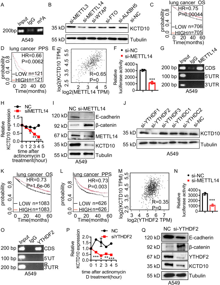
Lung cancer remains a critical global health concern, characterized by the highest incidence and mortality rates among all cancers. Due to its heterogeneity and complexity, the molecular mechanism underlying lung cancer occurrence and progression needs to be further investigated. KCTD10 has been implicated in malignant phenotypes of several tumors, but the role of KCTD10 in lung cancer remains largely unexplored. In this study, we found that KCTD10 expression is significantly reduced in lung cancer tissues, and overexpression of KCTD10 could inhibit lung cancer progression both in vitro and in vivo. Immunoprecipitation-mass spectrometry (IP-MS), co-immunoprecipitation (Co-IP), and ubiquitination assays revealed that the BTB domain of KCTD10 interacts with Armadillo repeat domains 1-9 of β-catenin and facilitates ubiquitin-dependent degradation of β-catenin via the K48-linked ubiquitin chains, followed by the downregulation of the β-catenin downstream target gene PD-L1. Notably, the combined treatment of KCTD10 overexpression with anti-PD-1 antibodies exhibited a synergistic effect in suppressing lung cancer progression and brain metastatic colonization in mice. In addition, vascular endothelial cell-specific knockout of Kctd10 (Kctd10flox/floxCDH5CreERT2/+) promoted lung cancer metastasis and tumor angiogenesis through β-catenin signaling. Finally, we identified METTL14- mediated N6-methyladenosine (m6A) modification within the coding sequence (CDS) region of KCTD10, which enhanced KCTD10 mRNA stability in a YTHDF2-dependent manner. These findings highlight KCTD10 as a critical regulator of lung cancer progression and the tumor microenvironment, suggesting its potential as a promising therapeutic target for lung cancer.

Keywords: KCTD10; M6A; PD-1; lung cancer metastasis; specific Kctd10 knockout; β-catenin.


Conflict of interest statement

The authors declare that the research was conducted in the absence of any commercial or financial relationships that could be construed as a potential conflict of interest.

Figures

Figure 1
Figure 1
Low expression of KCTD10 in lung cancer. (A) GEPIA database analysis of KCTD10 expression in normal lung tissues and lung cancer tissues. (B) Western blot analysis of KCTD10 expression in lung cancer cell lines and normal cell line Bears-2b. (C, D) IHC analysis of KCTD10 expression in human lung cancer tissues (n=80) and corresponding staining scores in different lung cancer grades. (E, F) IHC analysis of KCTD10 expression in LUAD (n=42) and LUSC (n=34). (G) Correlation between KCTD10 expression and overall survival in lung cancer patients, HR=0.65 (0.55-0.75), logrank P=1.2e-08. (H) Correlation between KCTD10 expression and post-progression survival of lung cancer patients, HR=0.64 (0.42-0.99), logrank P=0.042. (I) Correlation between KCTD10 expression and overall survival in LUAD patients, HR=0.62 (0.48-0.79), logrank P=0.00013. (J) Correlation between KCTD10 expression and disease free survival in LUAD patients, HR=0.41 (0.35-0.47), logrank P=0.042. *P < 0.05, **P < 0.01, ***P < 0.001.
Figure 2
Figure 2
Overexpression of KCTD10 inhibits lung cancer growth and metastasis in vitro and in vivo. (A) Fluorescence image showing the efficiency of lentiviral infection in A549 cells. (B) Western blot analysis confirming KCTD10 overexpression in A549 cells. (C) Colony formation assays demonstrating the effect of KCTD10 overexpression on cell growth. (D, E) Effects of KCTD10 on the weight and volume of subcutaneous A549 tumors (n=4/group). (F) Effects of KCTD10 on cell morphology of A549 subcutaneous tumors. (G, H) Transwell assays evaluating the effect of KCTD10 overexpression on A549 cell migration and invasion. (I, J) Effect of KCTD10 overexpression on lung colonization of A549 cells injected via the tail vein (n=4). 5x10⁵ A549 cells and KCTD10-overexpressing A549 cells were injected. (K, L) Western blot and IHC analysis of EMT-related gene expression. ***P< 0.001.
Figure 3
Figure 3
Interaction between KCTD10 and β-catenin proteins. (A) IP-MS and silver staining showing differential bands between anti-KCTD10 antibodies and IgG. (B) The top nine polypeptide scores of KCTD10-interacting proteins. (C) GEPIA database analysis of β-catenin expression in normal and lung cancer tissues. (D, E) IHC analysis of β-catenin expression and corresponding staining scores in different lung cancer grades (n=80). (F) IHC analysis of β-catenin in LUAD (n=42). (G) Correlation between KCTD10 and β-catenin expression in lung cancer. (H–K) Correlation between β-catenin expression and overall/post-progression survival of lung cancer patients. (H) HR=1.15 (1.02-1.29), logrank P=0.027. (I) HR=1.45 (1.08-1.97), logrank P=0.014. (J) HR=1.72(1.31-2.25), logrank P=6.3e-05. (K) HR=1.98 (1.29-3.05), logrank P=0.0015. (L) Western blot analysis of β-catenin expression and its downstream genes in A549 cells. *P < 0.05, **P < 0.01, ***P < 0.001.
Figure 4
Figure 4
KCTD10 promotes ubiquitination and degradation of β-catenin through the K48-ubiquitin chain. (A) Co-IP analysis demonstrating the interaction between KCTD10 and β-catenin proteins. (B) Representative schematic of KCTD10 and β-catenin protein domains. (C, D) Identification of the interacting regions between truncated KCTD10 and β-catenin proteins. (E) Degradation of β-catenin proteins after CHX treatment. (F) Effect of KCTD10 on β-catenin protein stability in the presence of MG132. (G, H) Fluorescence analysis and Western blots showing KCTD10-induced degradation of cytoplasmic β-catenin. (I–K) KCTD10 overexpression enhanced the ubiquitination of β-catenin. Myc-β-catenin was immunoprecipitated with rabbit polyclonal anti-β-catenin antibodies and these immunocomplexes were subjected to Western blotting with anti-ubiquitin antibodies to detect β-catenin-ubiquitin conjugates. In the ubiquitin constructs, R indicates that the corresponding lysine residue has been mutated to arginine, abolishing linkage at that site; O indicates that only the corresponding lysine residue remains intact, while all other lysines are mutated, allowing selective assessment of linkage through that specific site.
Figure 5
Figure 5
Overexpression of Kctd10 combined with anti-PD-1 therapy effectively suppresses lung tumor colonization. (A) Effect of Kctd10 expression on patient survival in high CD8 expression cohorts. (B) Combined therapeutic strategy for LLC tumors inoculated into C57BL/6 mice. (C, D) Construction and validation of stable LLC cell lines overexpressing Kctd10. (E, F) Effect of Kctd10 and anti-PD-1 therapy on lung tumor size and survival in LLC-bearing mice (n=4/group). (G) IHC analysis of β-catenin and Pd-l1 proteins expression following Kctd10 overexpression and anti-PD-1 treatment. (H) IF analysis of CD8a proteins expression following Kctd10 overexpression and anti-PD-1 treatment. *P < 0.05, **P < 0.01, ***P < 0.001.
Figure 6
Figure 6
Kctd10 in combination with anti-PD-1 therapy suppressed lung cancer brain metastasis. (A) Combined strategy for intracranial LLC metastatic tumor treatment in C57BL/6 mice. (B, C) Effects of Kctd10 and anti-PD-1 therapy on intracranial tumor sizes and survival in LLC-bearing mice (n=4/group). (D) IHC analysis of β-catenin and Pd-l1 proteins following Kctd10 overexpression and anti-PD-1 therapy. (E) IF analysis of CD8a proteins following Kctd10 overexpression and anti-PD-1 therapy. *P < 0.05, **P < 0.01, ***P < 0.001.
Figure 7
Figure 7
Endothelial Kctd10 knockout inhibits angiogenesis and metastatic phenotypes in lung tumors. (A) Construction strategy for Kctd10flox/floxCDH5CreERT2/+ mice. (B) Experimental strategy for inducible Kctd10 knockout and LLC cell injection in Kctd10flox/floxCDH5CreERT2/+ mice. Mice were treated by tamoxifen (75 mg/kg) for one week (n=5/group). (C, D) Images of lung cancer and corresponding HE staining in conditional knockout mice. (E, F) Immunofluorescence and IHC analysis of target gene expression in mouse lungs and lung tumor tissues. ***P < 0.001.
Figure 8
Figure 8
METTL14 and YTHDF2 mediates m6A modification of KCTD10 and enhances its mRNA stability. (A) MeRIP detecting m6A modification of KCTD10 CDS. (B) KCTD10 protein levels following m6A-related interfering RNAs. (C, D) Correlation between METTL14 expression and overall/post-progression survival in lung cancer patients using Kaplan-Meier Plotter survival analysis. (E) Correlation between METTL14 and KCTD10 expression in lung cancer using the GEPIA database. (F) Effects of METTL14 on the luciferase reporter activity of KCTD10. (G) RIP analysis detecting METTL14 binding to the predicted modification site of KCTD10. (H) qPCR analysis of KCTD10 RNA stability following METTL14 knockdown. (I) Western blot analysis of KCTD10 and related protein expression in A549 cells following METTL14 interference. (J) KCTD10 protein expression after m6A-related RNA interference. (K, L) Correlation between YTHDF2 expression and patient survival. (M) The GEPIA database analyzing the correlation between YTHDF2 and KCTD10 expression. (N) Effect of YTHDF2 knockdown on luciferase reporter activity of KCTD10. (O) RIP analysis of YTHDF2 binding to predicted modification sites of KCTD10 in A549 cells. (P) qPCR analysis of KCTD10 RNA stability following YTHDF2 knockdown. (Q) Western blot analysis of KCTD10 and downstream gene expression following YTHDF2 knockdown. *P < 0.05, **P < 0.01, ***P < 0.001.

Similar articles

References

    1. Khanmohammadi A, Aghaie A, Vahedi E, Qazvini A, Ghanei M, Afkhami A, et al. Electrochemical biosensors for the detection of lung cancer biomarkers: A review. Talanta. (2020) 206:120251. doi: 10.1016/j.talanta.2019.120251, PMID: - DOI - PubMed
    1. Sun D, Cao M, Li H, He S. Cancer burden and trends in China: A review and comparison with Japan and South Korea. Chin J Cancer Res. (2020) 32:129–39. C. W. doi: 10.21147/j.issn.1000-9604.2020.02.01, PMID: - DOI - PMC - PubMed
    1. Leiter A, Veluswamy RR. The global burden of lung cancer: current status and future trends. Nat Rev Clin Oncol. (2023) 20:624–39. doi: 10.1038/s41571-023-00798-3. W. JP., PMID: - DOI - PubMed
    1. Li Q, Lai Q, He C, Fang Y, Yan Q, Zhang Y, et al. RUNX1 promotes tumour metastasis by activating the Wnt/β-catenin signalling pathway and EMT in colorectal cancer. J Exp Clin Cancer Res. (2019) 38:334. doi: 10.1186/s13046-019-1330-9, PMID: - DOI - PMC - PubMed
    1. Zou Y, Lin X, Bu J, Lin Z, Chen Y, Qiu Y, et al. Timeless-Stimulated miR-5188-FOXO1/β-Catenin-c-Jun Feedback Loop Promotes Stemness via Ubiquitination of β-Catenin in Breast Cancer. Mol Ther. (2020) 28:313–27. doi: 10.1016/j.ymthe.2019.08.015, PMID: - DOI - PMC - PubMed