. 2025 Oct 28;121(13):2095-2112.
doi: 10.1093/cvr/cvaf146.

Mapping disease-specific vascular cell populations responsible for obliterative arterial remodelling during the development of pulmonary arterial hypertension

Affiliations

Mapping disease-specific vascular cell populations responsible for obliterative arterial remodelling during the development of pulmonary arterial hypertension

Nicholas D Cober et al. Cardiovasc Res. .

Abstract

Aims: Pulmonary arterial hypertension (PAH) is a lethal pulmonary vascular disease characterized by arteriolar pruning and occlusive vascular remodelling leading to increased pulmonary vascular resistance and eventually right heart failure. While endothelial cell (EC) injury and apoptosis are known triggers for this disease, the mechanisms by which they lead to complex arterial remodelling remain obscure. We employed multiplexed single-cell RNA sequencing at multiple timepoints during the onset and progression of disease in a model of severe PAH to identify mechanisms involved in the development of occlusive arterial lesions.

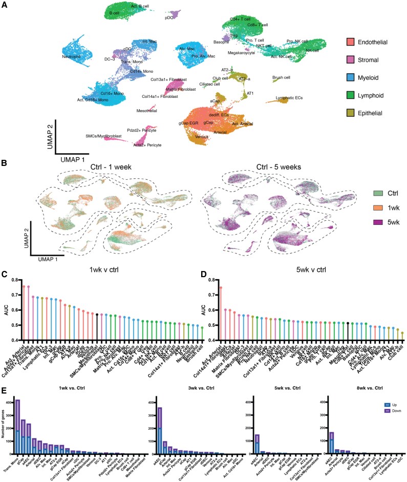
Methods and results: Single-cell transcriptional analysis resolved 44 global lung cell populations, with widespread early transcriptomic changes at 1 week affecting endothelial, stromal, and immune cell populations. In particular, two EC clusters were greatly expanded during PAH development and were identified as being disease specific: a relatively de-differentiated (dD) EC population that was enriched for Cd74 expression while exhibiting a loss of endothelial identity; and an activated arterial EC (aAEC) population that uniquely exhibited persistent differential gene expression throughout PAH development consistent with a growth regulated state. dDECs were primed to undergo endothelial-mesenchymal transition as evidenced by reduced activity of master EC transcription factors, Erg and Fli1, and further supported by RNA velocity analysis showing vectors leading to fibroblast clusters. Of note, aAECs exhibited high expression of Tm4sf1, a gene implicated in cancer cell growth, that was also expressed by a smooth muscle (SM)-like pericyte cluster, and were highly localized to regions of arterial remodelling in both the rat model and PAH patients, contributing to intimal occlusive lesions and SM-like pericytes forming bands of medial muscularization.

Conclusion: Together these findings implicate disease-specific vascular cells in PAH progression and suggest that TM4SF1 may be a novel therapeutic target for arterial remodelling.

Keywords: Endothelial cells; Pericytes; Pulmonary arterial hypertension; Vascular remodelling.


Conflict of interest statement

Conflict of interest: none declared.

Figures

Graphical Abstract
Graphical Abstract
Figure 1
Figure 1
Characterization of SU/CH model of PAH. (A) Timeline of the SU/CH model and relevant end of study sampling. (B) RVSP. (C) RVH (RV/LV + S). (D) Representative H&E histological images showing progression of arterial remodelling associated during development of PH. (E) Representative image of microCT of lung arterial angiograms during the development of PH. (F) Summary of quantitative analysis of arterial volumes by microCT showing the greatest reduction in arterioles under 150 µm in diameter. (G) Percent volume loss relative to healthy control in arteries under 200 µm in diameter. (H) Relationship between arterial volume (<500 µm diameter) and RVSP during PH development. Data represented as mean ± SEM, n = 6–12. A one-way ANOVA was used in Panels (B), (C), and (G) and a two-way ANOVA in Panel (F), with Tukey post hoc correction for multiple comparisons. *P < 0.05 for 3-, 5-, and 8-week SU/CH vs. control, †P < 0.05 for 3-, 5-, and 8-week vs. 1-week, ‡P < 0.05 for 5- and 8-week vs. 3-week, and #P < 0.05 for 0.01 for all timepoints vs. control. Scale bar represents 100 μm.
Figure 2
Figure 2
Multiplexed single-cell transcriptomic atlas of lung cells during PAH progression. (A) 44 unique lung clusters were identified and integrated into the global UMAP. (B) Global UMAPs coloured by timepoint with control (green) compared with 1 week (orange) and 5 weeks (purple). Cell prioritization was performed using a machine learning algorithm to identify clusters that were most affected between 1 week and control (C) and 5 weeks and control (D). At 1 week major transcriptomic changes indicated by an increase in area under the curve were seen in many populations with the greatest increases in aAECs and Acta2+ pericytes. However, at 5 weeks there was a general reduction in transcriptomic changes, with the exception of the aAEC population. (E) Number of all non-zero DEGs in lung cell populations at 1-, 3-, 5-, and 8-week post-SU compared with control (healthy). High numbers of DEGs were seen in transitional macrophages and many EC clusters at 1 week, but only aAECs displayed a persistently high number of DEGs at later timepoints. Single-cell data were obtained from n = 4 animals per timepoint, multiplexed using unique barcodes, pooled and subjected to library construction using 10x Genomics.
Figure 3
Figure 3
Changes in EC populations during PAH progression. (A) UMAP representation of all endothelial populations at all sampled timepoints showing 8 distinct cell types and a proliferative cell node enlarged in the box. (B) Feature plots highlighting genes typically associated with distinct EC populations including: Aplnr (gCap), Apln (aCap), Dll4 (arterial), Cxcl12 [‘activated’ arterial (aAECs) and arterial], Slc6a2 (venous), and Mmrn1 (lymphatic). (C) Heatmap showing the top 5 DEGs distinguishing each EC population. (D) Feature plots showing the distribution of proliferation-related genes, MKi67, Cdca8, and Birc5, largely within the proliferative node, highlighted and enlarged in the box. (E) UMAP of cells present at each timepoint demonstrating transcriptomic shifts compared to control at 1, 3, 5, and 8 weeks. (F) Fold change of each EC populations relative to their control levels. Early and persistent increases were seen in the relative size aAECs and dDECs, whereas arterial and lymphatic ECs showed a later expansion. Single-cell data were obtained from n = 4 animals per timepoint.
Figure 4
Figure 4
Transcriptomic signature of disease-specific aAEC and dDEC populations. Volcano plots for all differential expressed genes between (A) aAECs and arterial ECs and (B) dDECs and gCap ECs including all timepoints. (C) Dot plot showing relative expression of genes of interest between all EC clusters. dDECs exhibited the lowest expression of Cldn5 and Cdh5, and the highest expression of Cd74, RT1Da, and Tpt1, whereas aAECs showed the highest expression of Tm4sf1, among other genes associated with an activated phenotype and low expression of typical arterial genes (Dll4 and Cxcl4). (D) Ingenuity pathway analysis (IPA) comparing aAECs and arterial ECs showing the top 15 significantly up- and down-regulated gene sets. aAECs exhibited up-regulation of gene sets associated with cancer cell proliferation, migration and invasion, with down-regulation of gene sets for angiogenesis, vascular development and apoptosis. (E) IPA comparing dD and gCap ECs showing the top 15 significantly up- and down-regulated gene sets, demonstrating up-regulation of gene sets in dDECs associated with inflammation and apoptosis and down-regulation of gene sets associated with vascular development and angiogenesis. Single-cell data were obtained from n = 4 animals per timepoint.
Figure 5
Figure 5
Changes in stromal cell populations during PAH progression. (A) UMAP representation of all stromal populations at all sampled timepoints showing 6 distinct cell types. (B) Feature plots highlighting genes typical of distinct stromal populations including: pericytes (Cspg4, Mcam, and Pdgfrb); fibroblasts (Pdgfra, Col13a1, and Col14a1); SMCs (Acta2); and myofibroblasts (Dcn). (C) Heatmap of top five DEGs distinguishing each stromal population. (D) UMAP portraying the presence of cells from each stromal population at each timepoint, showing a marked expansion of the pericyte populations by 3 weeks. (E) Fold change of each stromal population relative to their respective controls demonstrating a marked relative increase in the SM-like Acta2+ pericytes and less so the ‘classical’ pericyte population during PAH progression. (F) Volcano plot of all DEGs between SM-like pericytes and classical pericytes. (G) Dot plot showing relative expression of genes of interest between different pericyte populations. (H) GSEA with the top 15 up- and down-regulated gene sets between SM-like pericytes and classical pericytes. Single-cell data were obtained from n = 4 animals per timepoint.
Figure 6
Figure 6
Endothelial transcriptomic changes during onset and progression of PAH. Volcano plots showing all DEGs for aAECs vs. control at 1 week (A) and 5 weeks (B). (C) Heatmap showing the top 20 up- and down-regulated DEGs for aAECs at each timepoint relative to control. Three transcriptional patterns can be discerned: (i) peak increase at 1 week with partial normalization at later timepoints, (ii) persistent elevation at all timepoints, and (iii) transient or sustained reduction in gene expression. (D) IPA for aAECs comparing each timepoint to the control, showing the top up- or down-regulated disease or biofunction gene sets for each timepoint. Single-cell data were obtained from n = 4 animals per timepoint.
Figure 7
Figure 7
Tm4sf1 as a marker for aAECs and SM-like pericytes in arterial remodelling. (A) Violin plots showing Tm4sf1 the highest expression in aAECs, with lower levels in arterial and venous ECs, SM-like pericytes and mesothelial cells. Immunofluorescent staining for TM4SF1 (white), isolectin B4 (ECs, green), 3G5 (pericytes, red), or SMA (SMCs, red) in healthy lungs (B) or at 5 weeks in the SU/CH severe PAH model (C). (D) Immunofluorescent staining of lung sections from control subjects and PAH patients for TM4SF1 (green), co-stained for ECs (CD31, red). Scale bar represents 20 μm (B and C) and 50 μm (D).
Figure 8
Figure 8
RNA velocity analysis indicates EndMT. (A) RNA velocity analysis of endothelial and stromal cell populations using scVelo’s dynamical model and CellRank demonstrate velocity vectors from aAEC and arterial EC populations, going through the dDEC cluster to fibroblast populations. RNA velocity vectors are also seen between classical pericytes SM-like (Acta2+) pericytes. Inset highlights the latent time analysis which estimates cellular differentiation and shows that gCap, aCap, fibroblasts, and classical pericytes approximate a more terminal differentiated state, while aAECs, dDECs, and SM-like pericytes exhibit a more progenitor-like state. (B) EC subset UMAP showing the distribution of dDECs in red. (C) UMAP showing the distribution of typical EMT related TF activities in the EC populations. (D) Heatmap of the top 40 most variable TFs in the different EC populations. The insert highlights reduced activity of Erg and Fli1, ETS family TFs that control endothelial identity, within cluster 1 (dDECs) and increased activity of Nr2f2 and Zeb2, which have been implicated in EndMT. Single-cell data were obtained from n = 4 animals per timepoint.

Comment in

References

    1. Simonneau G, Montani D, Celermajer DS, Denton CP, Gatzoulis MA, Krowka M, Williams PG, Souza R. Haemodynamic definitions and updated clinical classification of pulmonary hypertension. Eur Respir J 2019;53:1801913. - PMC - PubMed
    1. Evans CE, Cober ND, Dai Z, Stewart DJ, Zhao Y-Y. Endothelial cells in the pathogenesis of pulmonary arterial hypertension. Eur Respir J 2021;58:2003957. - PMC - PubMed
    1. Hoeper MM, Badesch DB, Ghofrani HA, Gibbs JSR, Gomberg-Maitland M, McLaughlin VV, Preston IR, Souza R, Waxman AB, Grünig E, Kopeć G, Meyer G, Olsson KM, Rosenkranz S, Xu Y, Miller B, Fowler M, Butler J, Koglin J, de Oliveira Pena J, Humbert M. Phase 3 trial of sotatercept for treatment of pulmonary arterial hypertension. N Engl J Med 2023;388:1478–1490. - PubMed
    1. Cober ND, VandenBroek MM, Ormiston ML, Stewart DJ. Evolving concepts in endothelial pathobiology of pulmonary arterial hypertension. Hypertension 2022;79:1580–1590. - PubMed
    1. Schupp JC, Adams TS, Cosme C, Raredon MSB, Yuan Y, Omote N, Poli S, Chioccioli M, Rose K-A, Manning EP, Sauler M, DeIuliis G, Ahangari F, Neumark N, Habermann AC, Gutierrez AJ, Bui LT, Lafyatis R, Pierce RW, Meyer KB, Nawijn MC, Teichmann SA, Banovich NE, Kropski JA, Niklason LE, Pe’er D, Yan X, Homer RJ, Rosas IO, Kaminski N. Integrated single-cell atlas of endothelial cells of the human lung. Circulation 2021;144:286–302. - PMC - PubMed

MeSH terms