. 2025 Aug 28;11(1):415.
doi: 10.1038/s41420-025-02691-7.

HapA protease targets PAR-1/2 to modulate ERK signalling and reduce cancer cell viability

Affiliations

HapA protease targets PAR-1/2 to modulate ERK signalling and reduce cancer cell viability

David Tena-Chaves et al. Cell Death Discov. .

Abstract

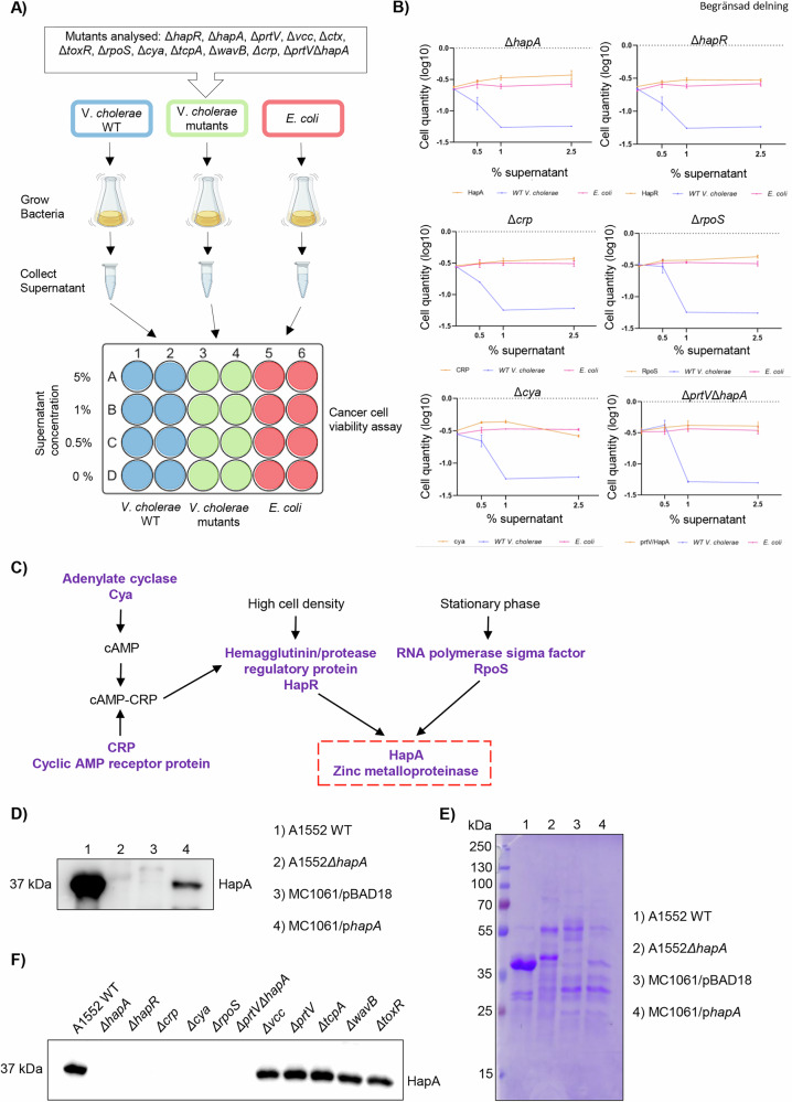
Recent studies reveal that Vibrio cholerae secretes virulence factors impacting host cell viability, though their effects on cancer cells remain unclear. However, the bacterial components and mechanisms influencing cancer cells remain largely unknown. This study investigated the effects of V. cholerae mutants lacking secreted proteins on carcinoma cells. We identified the hemagglutinin zinc-metalloprotease HapA as the main factor reducing cancer cell viability. HapA cleaves protease-activated receptors 1 and 2 on epithelial cancer cells at unique sites, unlike human proteases. This cleavage triggers an early and transient activation of the kinases MEK and ERK. Transient MEK and ERK activation initiates caspase 7, leading to apoptosis and reduced viability in epithelial cancer cells. Our findings underscore the significance of human protease-activated receptors as targets for bacterial protease HapA. Furthermore, we demonstrate that selective cleavage of PAR-1/2 by HapA adjusts MEK-ERK signalling dynamics, suggesting potential new avenues for the development of novel anticancer therapies. Understanding how pathogens like V. cholerae interact with cancer cells sheds light on potential mechanisms underlying cancer progression and suggests new therapeutic targets for cancer treatment.


Conflict of interest statement

Competing interests: AH provides advice to Novartis (clinical trial NeoLetTrib) and receives funding from Menarini pharma company. The other authors declare that there are no conflicts of interest. Ethics approval and consent to participate: We clarify that all experiments were conducted entirely in vitro. All methods were performed in accordance with the relevant guidelines and regulations. The study did not involve human participants, identifiable data or images, patient samples, volunteer-based research, or studies involving live vertebrates. No clinical or drug trials were conducted. Therefore, ethics approval and informed consent requirements do not apply.

Figures

Fig. 1
Fig. 1. Specific secreted proteins of V. cholerae impact the viability of cancer cells.
A Schematic representation of the cancer cell viability assay. B Viability assay in MCF-7 cells co-cultured with the supernatant from each of the 6 V. cholerae mutant strains with reverted effect, the wild-type V. cholerae, or the E. coli strain at concentrations of 0, 0.5, 1 and 2.5% for 24 h. Live cells were fixed and stained using crystal violet, and cell quantity was measured at 590 nm. Experiments were performed in triplicate, results were normalized applying Log10 and data are presented as mean ± SEM. (*) p-value < 0.05; (**) p-value < 0.01; (***) p-values < 0.001; (n.s) p-value > 0.05. C Bacterial regulatory pathway relating the proteins with a negative impact on cell viability in the assay. D A autodegradation HapA protein (37 KDa) detection from the supernatant of wild-type V. cholerae, its isogenic ΔhapA mutant, the E. coli K12 strain MC1061 habouring the pBAD18 vector, and MC1061 expressing HapA from V. cholerae. E Coomassie blue staining of the gel served as a loading control for the immunoblot shown in (D). F Western blot analysis showing HapA expression levels in supernatants isolated from A1552 wild-type and its isogenic mutants (ΔhapA, ΔhapR, Δcrp, Δcya, ΔrpoS, ΔprtVΔhapA, Δvcc, ΔprtV, ΔtcpA, ΔwavB, and ΔtoxR).
Fig. 2
Fig. 2. Mammalian PAR-1 and PAR-2 are cleaved by HapA.
A In silico analysis of consensus sites for PAR-1 cleavage by eukaryotic and prokaryotic proteases. B PAR-1 and PAR-2 reporter constructs carrying an alkaline phosphatase (AP) on their N-terminal end releases AP upon cleavage. The quantity of alkaline phosphatase present in the medium can be quantified by incubation with the colorimetric substrate 1-Step PNPP and measured at 405 nm. C, D PAR-1 and PAR-2 cleavage assay in MCF-7 (C) and MDA-MB-231 (D) cells. Cells transfected with the PAR-1 or PAR-2 reporter construct or the plasmid DNA without insertion were treated with the supernatant from the wild-type V. cholerae or from the ΔhapA mutant strain at a concentration of 0.5% for 20 min and 40 min. Alkaline phosphatase activity following PAR cleavage was quantified by incubation with the colorimetric substrate 1-Step PNPP and measured at 405 nm. The experiments were performed in triplicates; results were normalized applying Log10 and data are presented as mean ± SEM. (****) p-value < 0.0001. E PAR-1 cleavage assay in MCF7 cells transfected with the PAR-1 reporter construct or the plasmid DNA without insertion were treated with the supernatants from the wild-type V. cholerae, ΔhapA mutant strain, E. coli MC1061 expressing HapA or E. coli MC1061 empty vector at a concentration of 0.5% for 20 min and 40 min. Alkaline phosphatase activity following PAR1 cleavage was quantified by incubation with the colorimetric substrate 1-Step PNPP and measured at 405 nm. The experiments were performed in triplicates; results were normalized applying Log10 and data are presented as mean ± SEM. (****) p-value < 0.0001.
Fig. 3
Fig. 3. HapA induces MEK and ERK phosphorylation via PAR1.
A Schematic illustration of PAR-1 activation of MEK/ERK signalling pathway. PAR-1 activation induces conformational changes in its transmembrane domains, which favour the interaction of the receptor with heterotrimeric G proteins. In its inactive state, the Gαi/o subunit is bound to guanosine diphosphate (GDP). Activated GPCRs triggers a conformational change in the Gαi/o subunit, promoting the exchange of GDP for GTP. This nucleotide exchange results in dissociation of the Gαi/o subunit from the βγ complex. Coupling to Gαi/o inhibits adenylate cyclase (AC), suppressing the formation of c-AMP, and thereby activating MAPK signalling. B MCF-7 cells were transfected with the PAR-1 reporter construct and were treated with the supernatant from the wild-type V. cholerae (A1552 WT) or from the ΔhapA mutant (A1552 ΔhapA) strain at a concentration of 0.5% for 0, 20 and 40 minutes. Cells were collected, and protein extracts were analysed by SDS-PAGE and western blot for the expression of ERK, p-ERK, MEK and p-MEK. Vinculin was used as a loading control. The bottom part of the figure provides quantitative analysis of the experiment in MCF-7 cells with visualized bands for ERK, p-ERK (C), MEK and p-MEK (D) normalized by loading control. The experiments were performed in triplicates. A representative experiment is shown. (*) p-value < 0.05; (**) p-value < 0.01; (n.s) p-value > 0.05.
Fig. 4
Fig. 4. Inhibiting ERK phosphorylation mitigates the suppression of cell viability.
A MCF-7 cells were transfected with PAR-1 reporter construct and treated with the supernatant from the wild-type V. cholerae or HapA mutant strain at a concentration of 0,5% for 0 min and 20 min, in presence of either vehicle (DMSO) or trametinib (0,2 μM). Cells were collected, and protein extracts were analyzed by SDS-PAGE electrophoresis and western blot techniques for the levels of phospho-ERK, ERK and PAR-1. Vinculin was used as a loading control. (B) The plot provides quantitative analysis of the experiment in (A), in MCF-7 cells with visualized bands for ERK, p-ERK. (*) p-value < 0.05; (**) p-value < 0.01; (n.s) p-value > 0.05. (C) PAR1 cleavage assay of MCF-7 cells transfected as in (A) and cultured with the supernatant from the wild-type V. cholerae or HapA mutant strain at a concentration of 0,5% for 0 min and 20 min, in the presence of either vehicle (DMSO) or trametinib (0,2 μM). Alkaline phosphatase activity following PAR-1 cleavage was quantified by incubation with the colorimetric substrate 1-Step PNPP and measured at 405 nm. Data are presented as mean ± SEM from triplicate experiments whereby (****) p-value < 0.0001. D Viability assay in MCF-7 cells co-cultured with the supernatant from wild-type V. cholerae or HapA mutant strain, in the presence and absence of trametinib (0,2 μM) for 20 min. Subsequently, both bacterial supernatant and trametinib were removed, and cells were cultured in fresh media for 24 h prior fixation and staining of cells using crystal violet. Cell viability was quantified by measuring absorbance of crystal violet at 590 nm. The experiments were performed in triplicates, results were normalized, and data are presented as mean ± SEM. (*) p-value < 0.05; (n.s) p-value > 0.05.
Fig. 5
Fig. 5. HapA triggers apoptosis in MCF-7 cells.
A Quantification of live and dead MCF-7 cell percentages using the Incucyte® Artificial Intelligence Cell Health Analysis Module with 10X objective, 24 h after a 20-min incubation with 0.5% supernatant from either wild-type V. cholerae or the ΔhapA mutant strain. (B) Images of Caspase 3/7 Activity in MCF-7 cells treated with 0,5% of supernatant from wild-type V. cholerae or the HapA mutant strain, in the presence and absence of trametinib (0,2 μM) for 20 min. The cells were mixed with 1:1000 Caspase 3/7 Green Dye, and the plate was inserted in the Incucyte® Live Cell Imager system with 10X objective. Images were captured every hour. C Graph that represents total integrated Caspase 3/7 activity measured by emission of fluorescence at 530 nm from (B). The experiments were performed in triplicates. A representative experiment is shown. Results were normalized, and data are presented as mean ± SEM. (****) p-value < 0.0001; (n.s) p-value > 0.05. D Model of PAR-1 activation and downstream effects on cancer cells by HapA protease.

References

    1. Trivanovic D, Pavelic K, Persuric Z. Fighting cancer with bacteria and their toxins. Int J Mol Sci. 2021;22:12980. - PMC - PubMed
    1. Punj V, Bhattacharyya S, Saint-Dic D, Vasu C, Cunningham EA, Graves J, et al. Bacterial cupredoxin azurin as an inducer of apoptosis and regression in human breast cancer. Oncogene. 2004;23:2367–78. - PubMed
    1. Yamada T, Goto M, Punj V, Zaborina O, Chen ML, Kimbara K, et al. Bacterial redox protein azurin, tumor suppressor protein p53, and regression of cancer. Proc Natl Acad Sci USA. 2002;99:14098–103. - PMC - PubMed
    1. Gao M, Zhou J, Su Z, Huang Y. Bacterial cupredoxin azurin hijacks cellular signaling networks: protein-protein interactions and cancer therapy. Protein Sci. 2017;26:2334–41. - PMC - PubMed
    1. Toh E, Baryalai P, Nadeem A, Aung KM, Chen S, Persson K, et al. Bacterial protein MakA causes suppression of tumour cell proliferation via inhibition of PIP5K1alpha/Akt signalling. Cell Death Dis. 2022;13:1024. - PMC - PubMed

LinkOut - more resources