. 2025 Aug 29;10(1):275.
doi: 10.1038/s41392-025-02369-7.

The deacetylases HDAC1/HDAC2 control JAK2V617F-STAT signaling through the ubiquitin ligase SIAH2

Affiliations

The deacetylases HDAC1/HDAC2 control JAK2V617F-STAT signaling through the ubiquitin ligase SIAH2

Al-Hassan M Mustafa et al. Signal Transduct Target Ther. .

Abstract

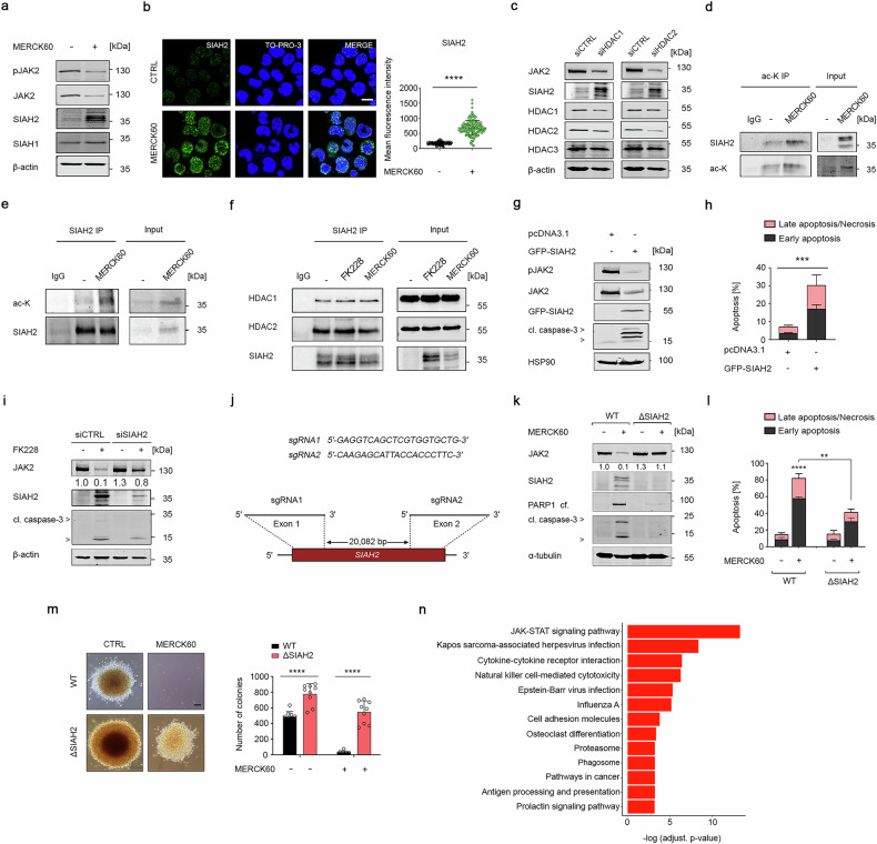
Epigenetic modulators of the histone deacetylase (HDAC) family control key biological processes and are frequently dysregulated in cancer. There is superior activity of HDAC inhibitors (HDACi) in patients with myeloproliferative neoplasms (MPNs) that carry the Janus kinase-2 point mutant JAK2V617F. This constitutively active tyrosine kinase activates signal-transducer-and-activator-of-transcription (STAT) transcription factors to promote cell proliferation and inflammatory processes. We reveal that the inhibition of HDAC1/HDAC2 with the clinically advanced HDACi romidepsin, the experimental HDACi entinostat and MERCK60, and genetic depletion of HDAC1/HDAC2 induce apoptosis and long-term growth arrest of primary and permanent MPN cells in vitro and in vivo. This treatment spares normal hematopoietic stem cells and does not compromise blood cell differentiation. At the molecular level, HDAC1 and HDAC2 control the protein stability of SIAH2 through acetylation. Genetic knockout experiments show that SIAH2 accelerates the proteasomal degradation of JAK2V617F in conjunction with the E2 ubiquitin-conjugating enzyme UBCH8. SIAH2 binds to the surface-exposed SIAH degron motif VLP1002 in the catalytic domain of JAK2V617F. At the functional level, SIAH2 knockout MPN cells are significantly less sensitive to HDACi. Global RNA sequencing verifies that JAK-STAT signaling is a prime target of SIAH2. Moreover, HDAC1 is an adverse prognostic factor in patients with acute myeloid leukemia (n = 150, p = 0.02), being a possible complication of MPNs. These insights reveal a previously unappreciated link between HDAC1/HDAC2 as key molecular targets, the still undefined regulation of cytoplasmic-to-nuclear signaling by HDACs, and how HDACi kill JAK2V617F-positive cells from MPN patients and mice with JAK2V617F in vitro and in vivo.


Conflict of interest statement

Competing interests: O.H.K. declares paid consultant activity for BASF, Ludwigshafen, Germany, and the patents “The use of molecular markers for the preclinical and clinical profiling of inhibitors of enzymes having histone deacetylase activity”, “Novel HDAC6 inhibitors and their uses”, and “Synthesis, pharmacology and use of new and selective FMS-like tyrosine kinase 3 (FLT3) inhibitors”. The BASF did not influence the study, and its products are not discussed in this work. All the other authors declare no conflict of interests.

Figures

Fig. 1
Fig. 1
Inhibition of class I HDACs induces apoptosis in JAK2V617F-positive cells. a HEL cells were treated with 5 nM FK228 for 24 h (-, untreated control cells; +, treated cells). Immunoblotting shows full-length (fl.) and cleaved (cf.) PARP1 and cleaved forms of caspase-3; β-actin was used as a loading control; kDa, kilo Daltons. b HEL cells were treated with 5 nM FK228 for 24–48 h, stained with annexin-V-FITC/PI, and analyzed for early (annexin-V-positive) and late (annexin-V/PI-positive) apoptosis by flow cytometry. Left shows representative flow cytometry scans; right shows bar graphs with statistical evaluations. c HEL cells were incubated with 5 nM FK228 and/or 50 μM z-VAD-FMK for 24 h. PARP1 was detected by immunoblotting; α-tubulin served as a loading control. d HEL cells were treated as described in (c) and subsequently stained with annexin-V-FITC/PI and analyzed for apoptosis. e HEL cells were treated as described in (c), fixed, subsequently stained with PI, and analyzed for subG1 fraction accumulation via flow cytometry. f HEL cells were treated with 5 nM FK228 for 24–48 h. The indicated proteins were revealed by immunoblotting; β-actin was used as a loading control. g HEL cells were treated with 5 nM FK228 for 24 h and/or proteasome inhibitors (50 nM bortezomib; 10 μM lactacystin) for 6 h before being harvested. Immunoblotting shows the indicated proteins. h HEL cells were treated as described in (g) and analyzed for apoptosis via flow cytometry. i HEL cells were incubated with 5 nM FK228 or 5 µM MS-275 for 24 h, washed, plated into methylcellulose medium, and tested for colony formation. Left, representative images are shown. Scale bar, 200 μm. Right, graph depicts the numbers of colonies. The data are presented as mean ± SD of at least three independent experiments. Statistical analyses (unpaired t-test; one-way ANOVA; two-way ANOVA; Bonferroni correction; ns not significant; p values are as follows: *P  < 0.05; **P  < 0.01; ***P  < 0.001; ****P  < 0.0001)
Fig. 2
Fig. 2
HDAC1 and HDAC2 protect JAK2V617F-positive cells from apoptosis. a HEL cells were incubated with 5 μM MERCK60 for 24 h. PARP1 and caspase-3 were detected by immunoblotting; β-actin, loading control. b HEL cells were treated as described in (a), stained with annexin-V-FITC/PI, and analyzed for apoptosis. c HEL cells were transfected with siRNAs against HDAC1 or HDAC2 for 48 h. The cells were stained with annexin-V-FITC/PI and analyzed for apoptosis. d GSEA analyses revealed the upregulation of proapoptotic gene signatures in HEL cells that were treated with 5 μM MERCK60 for 24 h, with a normalized enrichment score (NES) of 1.38 and an FDR q value < 0.05. e HEL cells were treated with 5 μM MERCK60 for 48 h, washed, plated in methylcellulose medium, and analyzed for colony formation after 14 days. Left, representative images are shown. Scale bar, 200 μm. Right, graph depicts the numbers of colonies. f Scheme depicting the experiment for which (g) shows the outcome; created with BioRender (https://BioRender.com). g MPN patient-derived JAK2-mutated PBMCs and HSPCs from healthy donors (HD) were treated with 0.5 or 1 μM MERCK60 or with DMSO as a control and plated in methylcellulose. The number of colonies and total number of cells were counted on day 10. Representative images are shown (upper panel). Scale bar, 200 μm. h Scheme depicting the experiment for which (i, j) show the outcome; created with BioRender (https://BioRender.com). i, j Hematopoietic stem and progenitor cells were isolated from the bone marrow of three mice with PV. Leukemic stem cells were enriched and tested for c-Kit and Sca-1 expression. The cells were treated with 5 µM MERCK60 for 24 h, washed, plated in methylcellulose, and analyzed for colony formation after 10 days; scale bar, 100 μm (i) or fixed, subsequently stained with PI, and analyzed for subG1 fraction accumulation via flow cytometry (j). k TCGA dataset analysis revealed that the overall survival of AML patients inversely correlates with the expression of HDAC1 mRNA. The data are presented as mean ± SD of three independent experiments. Statistical analyses (unpaired t-test; one-way ANOVA; two-way ANOVA; Bonferroni correction; ns not significant; *P  < 0.05; **P  < 0.01; ***P  < 0.001; ****P  < 0.0001)
Fig. 3
Fig. 3
Targeting HDAC1 and HDAC2 induces SIAH2-mediated degradation of JAK2V617F signaling. a HEL cells were incubated with 5 μM MERCK60 for 24 h. The cells were lysed, and the indicated proteins were analyzed by immunoblotting; β-actin served as a loading control. b HEL cells were treated with MERCK60 as described in (a), fixed, and stained for SIAH2. TO-PRO-3 was used to visualize the nuclei. The cells were examined via confocal laser scanning microscopy. Representative images are shown; n = 3; scale bar, 10 µm (left panel). The mean fluorescence intensities were measured with ImageJ software (right panel). c HEL cells were transfected with siRNAs against HDAC1 or HDAC2 for 48 h. Immunoblotting was used to verify the reduction in HDACs and the specificity of the siRNAs. Levels of JAK2V617F and SIAH2 were detected by immunoblotting; β-actin served as a loading control. d HEL cells were treated with 5 μM MERCK60 for 48 h. Immunoprecipitates with anti-pan-acetylated lysine (ac-K) antibody or rabbit preimmune serum (IgG) were analyzed for SIAH2 by immunoblotting. e K562 cells were treated as described in (d), and immunoprecipitates with an anti-SIAH2 antibody or mouse preimmune serum (IgG) were analyzed for pan-acetylated lysine (ac-K) by immunoblotting. f HEL cells were treated with 5 nM FK228 or 5 μM MERCK60 for 48 h. Immunoprecipitates with anti-SIAH2 antibody or mouse preimmune serum (IgG) were analyzed for interaction with HDAC1 and HDAC2 by immunoblotting. g HEL cells were transfected with pcDNA3.1 or pcDNA3.1-GFP-SIAH2 plasmids for 48 h. Immunoblotting was used to verify the expression of JAK2V617F, GFP-SIAH2, and cleaved caspase-3; HSP90, loading control. h HEL cells were transfected as described in (g), stained with annexin-V-FITC/PI and analyzed for apoptosis via flow cytometry. i HEL cells were transfected with siRNAs against SIAH2 for 48 h. Immunoblotting was used to verify the depletion of SIAH2. The levels of JAK2V617F and cleaved caspase-3 were detected by immunoblotting; β-actin served as a loading control. j Exon 1 and exon 2 of the human SIAH2 gene were disrupted by two gRNAs. k Wild-type (HELWT) and SIAH2 knockout HEL cells (HELΔSIAH2) were incubated with 5 µM MERCK60 for 48 h. Immunoblotting was used to verify the indicated proteins. α-tubulin served as a loading control. l Aliquots of cells from the experiments mentioned in (k) were stained with annexin-V-FITC/PI and analyzed for apoptosis. m HELWT and HELΔSIAH2 were treated as described in (k), washed, and analyzed for colony formation after 14 days. Left, representative images are shown. Scale bar, 100 μm. Right, graph depicts the numbers of colonies. n KEGG analysis shows the most enriched pathways in HELΔSIAH2 compared with HELWT cells, with an FDR q value < 0.05. The data are presented as mean ± SD of three independent experiments. Statistical analyses (unpaired t-test; one-way ANOVA; two-way ANOVA; Bonferroni correction; ns not significant; *P  < 0.05; **P  < 0.01; ***P  < 0.001; ****P  < 0.0001)
Fig. 4
Fig. 4
SIAH2 promotes the degradation of JAK2V617F through the VxP motif in its kinase domain. a Upper panel, illustration depicting the organization of JAK2 into the structural JH1-7 domains and the distribution of VxP motifs within its four functional domains. Illustration was created with BioRender (https://BioRender.com). Lower panel, 3D visualization of JAK2 showing VxP motifs (yellow) and the ubiquitin-binding domain (orange) via ChimeraX V1.3. b SIAH2 was expressed as a glutathione S-transferase (GST) fusion protein, GST-SIAH2. Samples were purified via Glutathione Sepharose 4B beads and analyzed for JAK2 binding by immunoblotting. c Immunoprecipitates with anti-SIAH2 antibody or mouse preimmune serum (IgG) were analyzed for pJAK2 and JAK2 by immunoblotting. HEL cells were incubated with 50 nM bortezomib for 6 h to prevent the proteasomal degradation of SIAH2-bound JAK2V617F. d VLP degron motif in the kinase domain determines JAK2 degradation by SIAH2. FLAG-tagged kinase domains of JAK2WT or mutant JAK2V1000G were coexpressed with GFP-SIAH2 in HEK293T cells. Immunoblotting verified the presence of the indicated proteins, and HSP90 served as a loading control. e HEL cells were transfected with siRNA against UBCH8 or noncoding siRNA. The cells were subsequently treated with 5 µM MERCK60 for 48 h. Immunoblotting was used to verify the expression of JAK2V617F and UBCH8; GAPDH served as a loading control. f HEL cells were transfected as described in (e), stained with annexin-V-FITC/PI and analyzed for apoptosis via flow cytometry. g UBCH8 was expressed as the glutathione S-transferase (GST) fusion protein GST-UBCH8. The samples were purified via Glutathione Sepharose 4B beads and analyzed for JAK2 binding by immunoblotting. h Illustration summarizing how HDAC1 and HDAC2 protect JAK2V617F from SIAH2-mediated proteasomal degradation. SIAH2 cooperates with UBCH8 and promotes polyubiquitylation and subsequent proteasomal degradation of JAK2V617F. Illustration was created with BioRender (https://BioRender.com). The data are presented as mean ± SD of three independent experiments. Statistical analyses (one-way ANOVA; two-way ANOVA; ns not significant; Bonferroni correction; **P  < 0.01; ***P  < 0.001; ****P  < 0.0001)
Fig. 5
Fig. 5
MERCK60 does not harm normal blood cells and is effective against MPN cells in vivo. a Fresh PBMCs were treated with increasing doses (1 µM, 2 µM, or 5 µM) of MERCK60 for 24 h. Staining for the survival markers annexin-V AF647 and FVD eFl780 was performed via flow cytometry. The isolated subtypes of cells were defined as follows: CD3-CD19+ as B-cells; CD3+ as T-cells; CD3-CD19-CD14+ as monocytes; CD3-CD19-CD1c+ as dendritic cells; CD3-CD19-CD56+ as natural killer (NK) cells. Illustration was created with BioRender (https://BioRender.com). b PBMCs were incubated with Dynabeads™ Human T-Activator CD3/CD28 (5 µl/ml cell suspension) for 24 h. Afterward, the cells were treated with MERCK60 (1 µM, 2 µM, or 5 µM) for 24 h. The cells were harvested and subjected to flow cytometry using FVD to discriminate live cells, CD3 as a T-cell marker, and CD25 to detect T-cell activation. c Scheme depicting the experimental setup. Whole bone marrow cells from JAK2V617F mice were treated ex vivo with either DMSO or 5 µM MERCK60 for 24 h. JAK2V617F cells (1 ×10⁵) were cotransplanted with 9 ×10⁵ cells into lethally irradiated Ly5.1 recipient mice. d Decrease in JAK2V617F-positive cells (CD45.2) in the peripheral blood (PB) at week 2 (left panel). Percentage of JAK2V617F-positive cells (CD45.2) at week 4 in the PB, bone marrow (BM), and spleen (SP) (middle panel). Pie charts depicting the engraftment of JAK2V617F-positive cells (%) after ex vivo treatment with MERCK60 or the DMSO control (right panel). The data represent mean ± SD of at least three independent experiments. Statistics (unpaired t-test; one-way ANOVA; ns not significant; P values are significant as shown)

References

    1. Perner, F. et al. Malignant JAK-signaling: at the interface of inflammation and malignant transformation. Leukemia39, 1011–1030 (2025). - PMC - PubMed
    1. Mustafa, A. M. & Krämer, O. H. Pharmacological modulation of the crosstalk between aberrant janus kinase signaling and epigenetic modifiers of the histone deacetylase family to treat cancer. Pharm. Rev.75, 35–61 (2023). - PubMed
    1. Philips, R. L. et al. The JAK-STAT pathway at 30: much learned, much more to do. Cell185, 3857–3876 (2022). - PMC - PubMed
    1. Chen, E. et al. JAK2V617F promotes replication fork stalling with disease-restricted impairment of the intra-S checkpoint response. Proc. Natl. Acad. Sci. USA111, 15190–15195 (2014). - PMC - PubMed
    1. Jayavelu, A. K. et al. Splicing factor YBX1 mediates persistence of JAK2-mutated neoplasms. Nature588, 157–163 (2020). - PubMed

MeSH terms

LinkOut - more resources