. 2025 Aug 28;8(1):1295.
doi: 10.1038/s42003-025-08713-5.

Inactivation mechanisms of Na+/ HCO 3 - cotransporter NBCe1 by phosphorylation

Affiliations

Inactivation mechanisms of Na+/ HCO 3 - cotransporter NBCe1 by phosphorylation

Han Wu et al. Commun Biol. .

Abstract

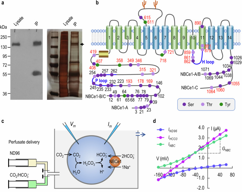
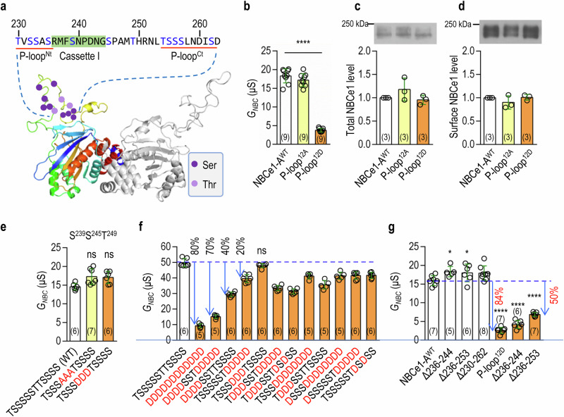
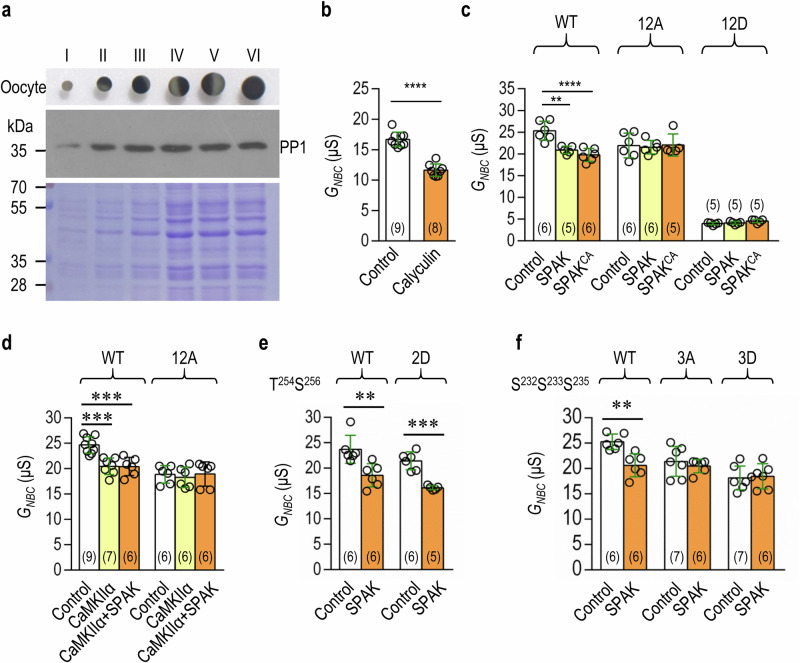
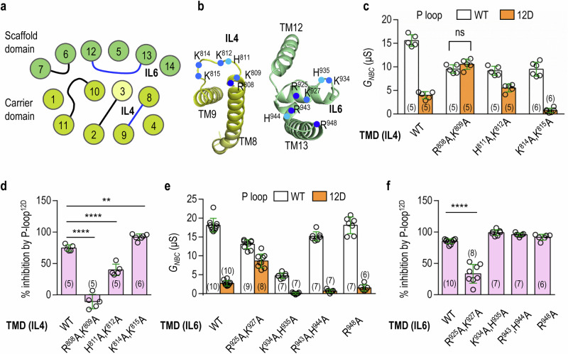
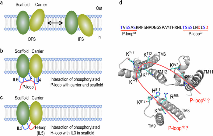
The solute carriers (SLC) superfamily comprises 66 families with more than 450 members. The Na+/ HCO 3 - cotransporter NBCe1 (SLC4A4) of SLC4 family plays critical roles in intracellular pH regulation and transepithelial transport of fluid and electrolytes. Here, we explored the structural mechanisms of NBCe1-A regulation by two phosphorylation modules: P-loop in the amino-terminal domain and H-loop in the transmembrane domain. Mimic-phosphorylation of P-loop or H-loop substantially decreases NBCe1-A activity. Inhibition of NBCe1 by P-loop is abolished by mutations to specific basic residues in the fourth intracellular loop (IL4) in the carrier domain and IL3/IL6 in the scaffold. Inhibition by H-loop is abolished by specific mutations to IL3. We conclude that: (1) P-loop inactivates NBCe1-A by binding to the carrier and the scaffold; (2) H-loop blocks NBCe1-A by interacting with IL3 in the scaffold. Our findings have implications for studying the structural mechanisms for the regulation of other SLCs by phosphorylation.


Conflict of interest statement

Competing interests: The authors declare no competing interests.

Figures

Fig. 1
Fig. 1. Phosphosite identification and activity measurement of NBCe1.
a Western blotting (left) and silver staining (right) of NBCe1 preparations. Shown here is an example of NBCe1 isolated from the lysate of rat brain by immunoprecipitation (IP). The band indicated by arrow in the right panel was excised for the identification of phosphosites in NBCe1. b Topological model showing the distribution of all known phosphosites in NBCe1. See Table S1 for the source of the phosphosites. Shown in blue lines are the two phosphorylation modules P-loop in NTD and H-loop (the fifth intracellular loop IL5) in TMD. Green cylinders represent the transmembrane α helices (TM) in the carrier domain. Blue cylinders indicate the TMs in the scaffold domain. The numbering of residues is based on human NBCe1. The numbering of the residues in the conserved regions is according to human NBCe1-B (NCBI accession no. NP_001091954.1). The numberings in red indicate the novel phosphosites identified in the present study. Also see Table S1. c Diagram showing the perfusion of Xenopus oocytes for electrophysiology recordings. The oocytes were first perfused with nominally HCO3-free ND96 and then with a solution containing 5% CO2/33 mM HCO3. d Representative I-V curves of an oocyte expressing NBCe1-A. IND96 (blue) represents the gross current recorded in ND96. IHCO3 (purple) represents the gross current in CO2/HCO3 solution. The differential current (INBC, green) represents the net HCO3-dependent component mediated by NBCe1-A. The slope conductance (GNBC) of INBC between ±40 mV was used as an index for NBCe1 activity.
Fig. 2
Fig. 2. Mimic-phosphorylation of P-loop decreases NBCe1-A activity.
a 3D structure model of the NTD of NBCe1. The model of NBCe1 NTD was generated by homology modelling with the structure of NDCBE NTD (PDB: 5JHO) as template. P-loop (residues 230 − 262), which belongs to a large loop structure that is intrinsically-disordered in the NTD of NBCe1, contains 12 potential phosphosites (residues Ser and Thr) with two clusters in P-loopNt and P-loopCt. Highlighted in green is the optional Cassette I. b Mimic-phosphorylation of P-loop (P-loop12D) inhibits NBCe1-A activity. P-loop12A: 12 phosphosites replaced by Ala. P-loop12D: 12 phosphosites replaced by Asp. P-loop12A and P-loop12D have no significant effect on the relative expression levels of total (c) and surface (d) NBCe1-A in Xenopus oocytes. See Fig. S4a for full blots. e Mimic-phosphorylation of the middle portion of P-loop has no effect on NBCe1-A. f Maximal inhibition on NBCe1-A requires mimic-phosphorylation of both P-loopNt and P-loopCt. g Effect of P-loop deletions on NBCe1-A activity. Open bars: deletions based on NBCe1-AWT. Orange bars: deletions based on P-loop12D. Data are presented as mean ± SD. The numerals in the parentheses on the bars indicate the number of individual oocytes for activity data or the number of western blottings with independent pools of oocytes for expression data. One-way ANOVA followed by post hoc Dunnett’s multiple comparisons was used for statistical analysis. *p < 0.05; ****p < 0.0001, compared with NBCe1-AWT. ns not significant.
Fig. 3
Fig. 3. Overexpression of protein kinases decreases NBCe1-A activity.
a Expression of endogenous protein phosphatase PP1 in Xenopus oocytes. Upper panel: oocytes of stages I − VI. Middle panel: Western blotting for PP1. Bottom a parallel gel stained with Coomassie brilliant blue to show the loading. See Fig. S4b for full blots. b Administration of PP1 inhibitor Calyculin inhibits NBCe1-A activity in oocytes. c, d Overexpressing SPAK and/or CaMKIIα has no effect on the activities of P-loop12A or P-lop12D. SPAKCA: constitutively-active SPAK containing mimic-phosphorylation mutations T243E and S383D. e SPAK significantly decreases the activity of NBCe1-A with mimic-phosphorylation at T254 and S256. f SPAK has no effect on NBCe1-A with mimic-(de)phosphorylation at S232, S233, and S235. The numerals in the parentheses on the bars indicate the number of individual oocytes. One-way ANOVA followed by post-hoc Dunnett’s multiple comparisons was used for statistical analyses for a, b. Student’s t-test was used for c, d, f. **p < 0.01; ***p < 0.001; ****p < 0.0001.
Fig. 4
Fig. 4. Mutation to the basic residues in IL4 and IL6 in the TMD abolishes the inhibition of NBCe1-A by P-loop.
a Diagram showing the spatial arrangement of transmembrane α helices (TMs) in NBCe1 TMD (bottom view). Numbered circles represent the TMs. Yellow: TMs in the carrier domain. Green: TMs in the scaffold domain. b Localization of basic residues in IL4 and IL6. c Effect of mutations to basic residues in IL4 on the activity of NBCe1-A with either P-loopWT or P-loop12D. d Summary of the percentile inhibition of NBCe1-A by P-loop12D. The GNBC of individual oocytes contributed to the orange bars in panel c was subtracted from the mean of the corresponding control (open bar) and then divided by the mean of the control. The % difference represents the percentile inhibition of NBCe1-A by P-loop12D. e Effect of mutations to IL6 on the activity of NBCe1-A with either P-loopWT or P-loop12D. f Summary of percentile inhibition of NBCe1-A by P-loop12D. The figure was created based on the data contributed to e. Data are presented as mean ± SD. The numerals in the parentheses on the bars indicate the number of individual oocytes. ns not significant by student’s t-test. In d and f, one-way ANOVA followed by post-hoc Dunnett’s multiple comparisons was used for statistical analyses. **p < 0.01; ****p < 0.0001.
Fig. 5
Fig. 5. Mutation to the basic residues in IL3 in the TMD abolishes the inhibition of NBCe1-A by P-loop.
a Diagram showing the spatial arrangement of TMs in NBCe1 TMD (bottom view). Yellow: TMs in the carrier domain. Green: TMs in the scaffold domain. b Localization of basic residues in IL4 in the carrier (yellow), and IL3 and IL6 in the scaffold (green). c Effect of mutations to the basic residues in IL3 on the activity of NBCe1-A with either P-loopWT or P-loop12D. d Summary of percentile inhibition of NBCe1-A by P-loop12D. The figure was created based on the data contributed to c. e Proposed model for the mechanism of NBCe1-A inhibition by phosphorylation of P-loop. Data are presented as mean ± SD. The numerals in the parentheses on the bars indicate the number of individual oocytes. ns, not significant by two-tailed student’s t-test. In d, one-way ANOVA followed by post-hoc Dunnett’s multiple comparisons was used for statistical analyses. ****p < 0.0001.
Fig. 6
Fig. 6. Mimic-phosphorylation of H-loop inhibits NBCe1-A by interaction with IL3.
a Diagram showing the arrangement of TMs in NBCe1 TMD (bottom view). Yellow: TMs in the carrier domain. Green: TMs in the scaffold domain. H-loop is shown in red. IL3 is shown in blue. b Side view of NBCe1 TMD (partial). Note that H-loop is spatially close to TM6-IL3. Mutations to H-loop have no significant effect on the relative expression levels of total (c) and surface (d) NBCe1-A in oocytes. H-loop3A: T859/T861/S862 substituted with Ala. H-loop3D: T859/T861/S862 substituted with Asp. See Fig. S4c for full blots. e Mimic-phosphorylation of H-loop inhibits NBCe1-A activity. f, g Effect of mutations to IL3 on the activity of NBCe1-A with either H-loopWT or H-loop3D. h Summary of percentile inhibition of NBCe1-A by H-loop. The figure was created based on the data contributed to f and g. i Proposed model for the mechanism of NBCe1-A inhibition by phosphorylation of H-loop. Data are presented as mean ± SD. The numerals in the parentheses on the bars indicate the number of individual oocytes for activity data or the number of western blottings with independent pools of oocytes for expression data. ns, not significant by two-tailed student’s t-test in f and g. One-way ANOVA followed by post hoc Dunnett’s multiple comparisons was used for statistical analyses in h. **p < 0.01; ****p < 0.0001.
Fig. 7
Fig. 7. Summary of hypothetical models for the structural mechanisms of NBCe1 inactivation by phosphorylation of P-loop and H-loop.
a Conformation transition of NBCe1 during ion translocation. NBCe1 employs an elevator-like mechanism for ion translocation. The carrier domain (yellow) slides back and forth along the scaffold (green), causing the transition of NBCe1 between the outward-facing state (OFS) and the inward-facing state (IFS). b Inactivation of NBCe1-A by P-loop. Upon phosphorylation, P-loop simultaneously binds to IL4 in the carrier and IL3 and IL6 in the scaffold. This long-distance interaction disrupts the sliding of the carrier along the scaffold, causing NBCe1-A blockade. c Inactivation of NBCe1-A by H-loop. Upon phosphorylation, H-loop in the carrier binds to the IL3 in the scaffold. This local interaction interferes with the sliding of the carrier, therefore causing NBCe1-A blockade. d Hypothetical orientation of P-loop in its interaction with the TMD. P-loop contains two clusters of phosphosites in P-loopNt and P-loopCt. It is presumed that, upon phosphorylation, P-loopNt binds to IL4 in the carrier, whereas P-loopCt binds to IL3 and IL6 in the scaffold. See discussion in the main text.

Similar articles

References

    1. Meixner, E. et al. A substrate-based ontology for human solute carriers. Mol. Syst. Biol.16, e9652 (2020). - PMC - PubMed
    1. Dvorak, V. & Superti-Furga, G. Structural and functional annotation of solute carrier transporters: implication for drug discovery. Expert Opin. Drug Discov.18, 1099–1115 (2023). - PubMed
    1. Lin, L., Yee, S. W., Kim, R. B. & Giacomini, K. M. SLC transporters as therapeutic targets: emerging opportunities. Nat. Rev. Drug Discov.14, 543–560 (2015). - PMC - PubMed
    1. Hediger, M. A., Clemencon, B., Burrier, R. E. & Bruford, E. A. The ABCs of membrane transporters in health and disease (SLC series): introduction. Mol. Asp. Med.34, 95–107 (2013). - PMC - PubMed
    1. Jardetzky, O. Simple allosteric model for membrane pumps. Nature211, 969–970 (1966). - PubMed

Substances

LinkOut - more resources