Review
. 2025 Aug 29;6(1):60.
doi: 10.1186/s43556-025-00305-3.

Damage-associated molecular patterns (DAMPs) in diseases: implications for therapy

Affiliations
Review

Damage-associated molecular patterns (DAMPs) in diseases: implications for therapy

Heya Lin et al. Mol Biomed. .

Abstract

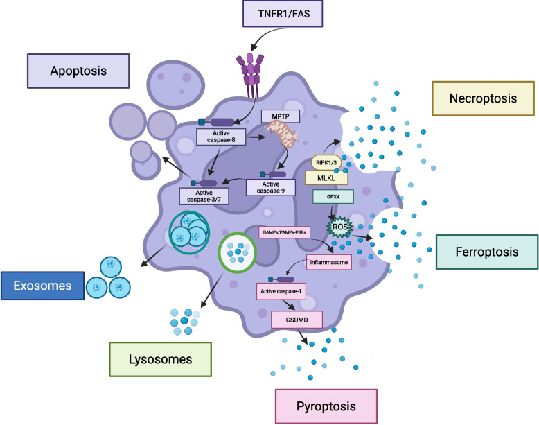
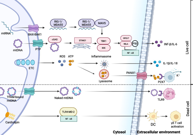
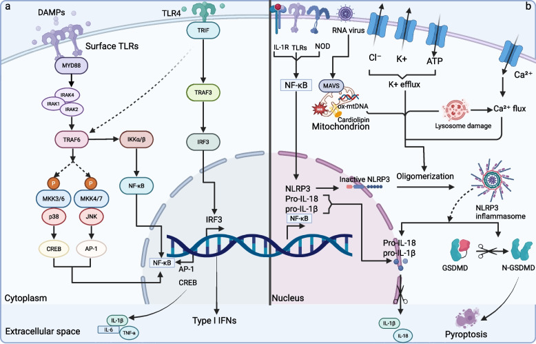
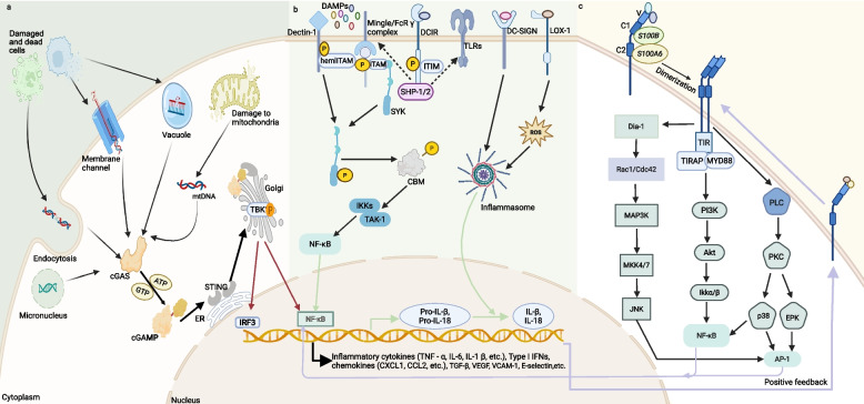
Damage-associated molecular patterns (DAMPs) are endogenous danger signal molecules released by damaged, stressed or dead cells that bind to pattern recognition receptors (PRRs), activating immune responses and inflammatory signaling pathways to play critical regulatory roles in various pathophysiological processes. This review classifies DAMPs into three major categories (protein-based, nucleic acid-based and mitochondria-derived) based on distinct molecular characteristics and biological functions, analyzing their structural features and functional differences. We systematically summarize current understanding of DAMP molecular transformation mechanisms, release pathways and recognition processes, with in-depth discussion of their pathological roles in major diseases including cancer, cardiovascular diseases and respiratory disorders. Particular emphasis is placed on the molecular recognition mechanisms between DAMPs and PRRs (TLRs, NLRs, CLRs and RAGE), and the disease regulatory networks formed by activated key signaling pathways (NF-κB, MAPK, inflammasomes and cGAS-STING). Current DAMP/PRR-targeted therapeutic strategies are comprehensively reviewed, including: modulating cell death pathways to reduce DAMP release, neutralizing DAMP activity using monoclonal antibodies, developing small-molecule inhibitors to block signaling pathways, and employing enzymatic degradation or gene silencing technologies for precise intervention. While showing promise in inflammatory and cancer disease models, these approaches face clinical translation challenges including DAMP molecular heterogeneity, inefficient drug delivery systems, and the complexity of multi-target synergistic mechanisms. Potential solutions involving nanoparticle delivery systems, AI-driven personalized treatment optimization and gene editing technologies are discussed. This review aims to provide references for developing novel therapeutics targeting the DAMP/PRR signaling axis, potentially opening new treatment avenues for cancer, neurodegenerative diseases, cardiovascular diseases and inflammatory disorders.

Keywords: Damage-associated molecular patterns; Immune response; Pathways; Pattern recognition receptors; Therapeutic strategies.


Conflict of interest statement

Declarations. Ethics approval and consent to participate: Not applicable. This scoping review did not involve human participants or animal subjects. Competing interests: The authors have no relevant financial or non-financial interests to disclose.

Figures

Fig. 1
Fig. 1
The source of DAMP. DAMP is mainly derived from innate immune cells including monocytes, macrophages, dendritic cells, neutrophils, mast cells, natural killer cells, eosinophils and others. Adaptive immune cells, including B cells and T cells. Non-immune cells, including endothelial cells, epithelial cells and fibroblasts, etc. DAMP, as a signalling molecule, interacts with these cells and plays an important role in initiating the immune response and regulating the immune response, thus bridging the gap between cellular damage and immune response
Fig. 2
Fig. 2
The release mechanisms of DAMPs. The different release mechanisms of DAMPs can be broadly classified into two categories:passive release mainly due to cell death and active release from living cells represented by cytotoxicity. Cell death modes include necrosis, pyroptosis, apoptosis, and Ferroptosis
Fig. 3
Fig. 3
Redox states of HMGB1. The three redox states of HMGB1 differ in structure and activity. The reduced form contains sulfhydryl groups on all three cysteine residues, and it is this form that is able to bind to RAGE and activate inflammatory pathways. The disulfide form contains a disulfide bond between the cysteine residues at positions 23 and 45, and this bond increases its stability. The final inactivated form is known as the oxidised form and has sulphonated cysteines at all three positions
Fig. 4
Fig. 4
Mitochondria-derived DAMPs. Mitochondrial RNA (mtRNA) is released through the BAX-BAK1 channel, activating RIG—1/MDA5 and subsequently signalling via MAVS. Mitochondrial DNA (mtDNA) is released via PTP, recognised by cGAS and activates STING1, which in turn acts on TBK1 and IKK, ultimately affecting IRF3/7 and NF-κB, and contributing to INF-β/IL-6 production. ROS and ATP produced by the mitochondrial electron transport chain (ETC) act on inflammatory vesicles to promote IL-1β/IL-18 release. In addition, lysosome-associated PANX1 and P2X7 are involved in K+ efflux and apoptosis. After cell death, TFAM-bound mtDNA and naked mtDNA activate TLR9 to mediate the inflammatory response, and cardiolipin activates TLR4-MD-2 and NF-κB, ultimately triggering dendritic cell (DC) and γδ T cell activation
Fig. 5
Fig. 5
DAMPs and immune signal transduction. The left a figure demonstrates that TLRs bind to ligands to form homo-/heterodimers, and after dimerization, their TIR domains recruit the adaptor protein TIRAP, which binds to the junction protein MyD88 and catalyzes the kinase of IPAKs, activates TRAF6, which activates the IKK complex and activates NF-κB, which enters the nucleus and induces the up-regulation of transcription, and promotes the transcription of pro-inflammatory cytokine (e.g., TNF-α, IL-1β, IL-6) transcription. Meanwhile, TRAF6 also catalyzes MKKs, activates the JNK/p38 pathway, and induces AP-1 transcription, which in turn mediates RNA regulation.TLR3 and TLR4 activate the TRAF3-IRF3 axis through the TRIF-dependent pathway to induce type I interferon production. The right b panel shows the classical activation pathway of NLRP3 inflammasome: after sensing signals from mitochondrial ROS, ionic currents (K+ efflux, Ca2+ efflux), and lysosomal damage by the intracellular NLRP3 receptor, it assembles with ASC proteins to form an inflammasome that cleaves and activates caspase −1. Caspase −1, on the one hand, cleaves pro—IL—1β, on the other. Caspase-1, on the one hand, cleaves pro-IL-1β and pro-IL-18 as mature cytokines that mediate the inflammatory cascade; on the other hand, it cleaves GSDMD proteins to form N-GSDMD, which drives cellular pyroptosis and releases inflammatory contents to amplify the immune response
Fig. 6
Fig. 6
DAMPs-mediated proinflammatory signal transduction. This schematic illustrates the activation mechanisms of three innate immune signaling pathways: a In the cGAS-STING pathway, cytosolic dsDNA activates cGAS to produce cGAMP, which induces STING translocation and subsequently activates the IRF3/NF-κB pathway via TBK1, leading to the production of pro-inflammatory cytokines; b In the DAMP-sensing pathway, activating receptors (e.g., Dectin-1) engage the SYK/CBM complex to trigger the TAK1-IKKs-NF-κB cascade, promoting pro-IL-1β transcription, while inhibitory receptors (e.g., DCIR) suppress SYK activity through SHP-1/2, and the NLRP3 inflammasome cleaves pro-IL-1β to generate mature IL-1β; c In the RAGE signaling pathway, ligand-bound RAGE activates NF-κB via PI3K-AKT-IKK or AP-1 via Rac1-MAP3K-JNK, thereby releasing cytokines/MMPs, while HMGB1-RAGE amplifies NF-κB/AP-1 signaling through the PLC-PKC axis, establishing a pro-inflammatory feedback loop
Fig. 7
Fig. 7
Role of DAMPs in tumor immunity. Immunomodulation and inflammatory response in the tumor microenvironment: immunogenic cell death DAMPs such as HSP, HMGB1, mtDNA, etc., activate receptors such as TLRs, RAGE, NLRs, etc., which in turn triggers pathways such as MAPK, NLRP3 inflammasome NF -κB, etc., to promote the release of pro-inflammatory cytokines and recruitment of immune cells. At the same time, immunosuppressive cells are activated and expanded to secrete cytokines such as IL-10 and TGF-β, creating a complex balance between pro- and anti-tumor. Inflammatory response leads to the destruction of the epithelial barrier and the degradation of the extracellular matrix through MMPs, which ultimately promotes the growth, proliferation and metastasis of tumor cells
Fig. 8
Fig. 8
DAMPs—driven cardiac inflammatory injury. Signaling and regulatory mechanisms of DAMPs in cardiovascular pathological processes: hypoxia, overload and hypertension promote DAMPs in cardiomyocytes. these molecules activate downstream pathways via receptors such as cGAS-STING, NOD, TLRs: MyD88 activates NF-κB, triggering inflammatory responses and recruiting immune cells; TRIF activates IRF. meanwhile. cfDNA activates NLRP3 inflammasome, generating ROS and exacerbating programmed cell death. At the same time, cfDNA activates NLRP3 inflammatory vesicles and generates ROS, promoting programmed cell death and exacerbating myocardial fibrosis. On the right side, DAMPs activated platelets to promote thrombosis and vascular calcification, activated neutrophils to produce reactive oxygen species and induced NETosis, and acted on endothelial cells and mesenchymal stromal cells to participate in angiogenesis and foam cell formation
Fig. 9
Fig. 9
DAMPs—mediated multi—organ inflammatory injury. Schematic diagram of the mechanism of DAMPs in multi-organ pathological processes: This figure demonstrates the mechanism by which molecules such as S100, HMGB1, and HSP activate signaling pathways such as NF-κB, MAPK, and NLRP3 through receptors such as TLRs, NLRs, and RAGE, triggering the release of pro-inflammatory cytokines and recruitment of immune cells, which then mediate the mechanism of multi-organ pathological changes. In the lungs, it can induce acute respiratory distress syndrome, pneumonia, lung injury, pulmonary edema and pulmonary fibrosis; in the liver, it activates hepatic stellate cells, leading to hepatic fibrosis, hepatic failure, fatty liver and cirrhosis, etc.; and in the kidneys, it involves renal fibrosis, nephritis, acute renal injury, and renal vascular lesions. In addition, the effects of neutrophil extracellular traps on platelets and renal vasculature, as well as the effects of IL-22 on podocytes, are shown, presenting a comprehensive picture of the complex regulatory network of these molecules in multi-organ pathologic processes

Similar articles

References

    1. Janeway CA. Approaching the asymptote? Evolution and revolution in immunology. Cold Spring Harb Symp Quant Biol. 1989;54:1–13. 10.1101/SQB.1989.054.01.003. - PubMed
    1. Matzinger P. Tolerance, danger, and the extended family. Annu Rev Immunol. 1994;12(1):991–1045. 10.1146/annurev.iy.12.040194.005015. - PubMed
    1. Seong S-Y, Matzinger P. Hydrophobicity: an ancient damage-associated molecular pattern that initiates innate immune responses. Nat Rev Immunol. 2004;4(6):469–78. 10.1038/nri1372. - PubMed
    1. Kim YS, Park HJ, Park JH, Hong EJ, Jang G-Y, Jung ID, et al. A novel function of API5 (apoptosis inhibitor 5), TLR4-dependent activation of antigen presenting cells. OncoImmunology. 2018;7(10): e1472187. 10.1080/2162402X.2018.1472187. - PMC - PubMed
    1. Park HJ, Jang G-Y, Kim YS, Park JH, Lee SE, Vo M-C, et al. A novel TLR4 binding protein, 40S ribosomal protein S3, has potential utility as an adjuvant in a dendritic cell-based vaccine. J Immunother Cancer. 2019;7(1):60. 10.1186/s40425-019-0539-7. - PMC - PubMed

LinkOut - more resources