. 2025 Aug 29;44(1):258.
doi: 10.1186/s13046-025-03508-2.

PD-1+ NK cell subsets in high grade serous ovarian cancer: an indicator of disease severity and a target for combined immune-checkpoint blockade

Affiliations

PD-1+ NK cell subsets in high grade serous ovarian cancer: an indicator of disease severity and a target for combined immune-checkpoint blockade

Marco Greppi et al. J Exp Clin Cancer Res. .

Abstract

Background: Ovarian cancer (OC) is the fifth leading cause of cancer-related death among women, with High-Grade Serous Ovarian Carcinoma (HGSC) representing the most aggressive and prevalent subtype. Despite promising results in other malignancies, immune checkpoint blockade has shown limited efficacy in HGSC, highlighting the need for alternative immunotherapeutic targets.

Methods: We conducted an integrated analysis combining multiparametric flow cytometry, RNA sequencing, multiplex immunohistochemistry, and functional assays to characterize NK cells isolated from peripheral blood, peritoneal fluid, primary tumor tissue, and metastases in 60 HGSC patients.

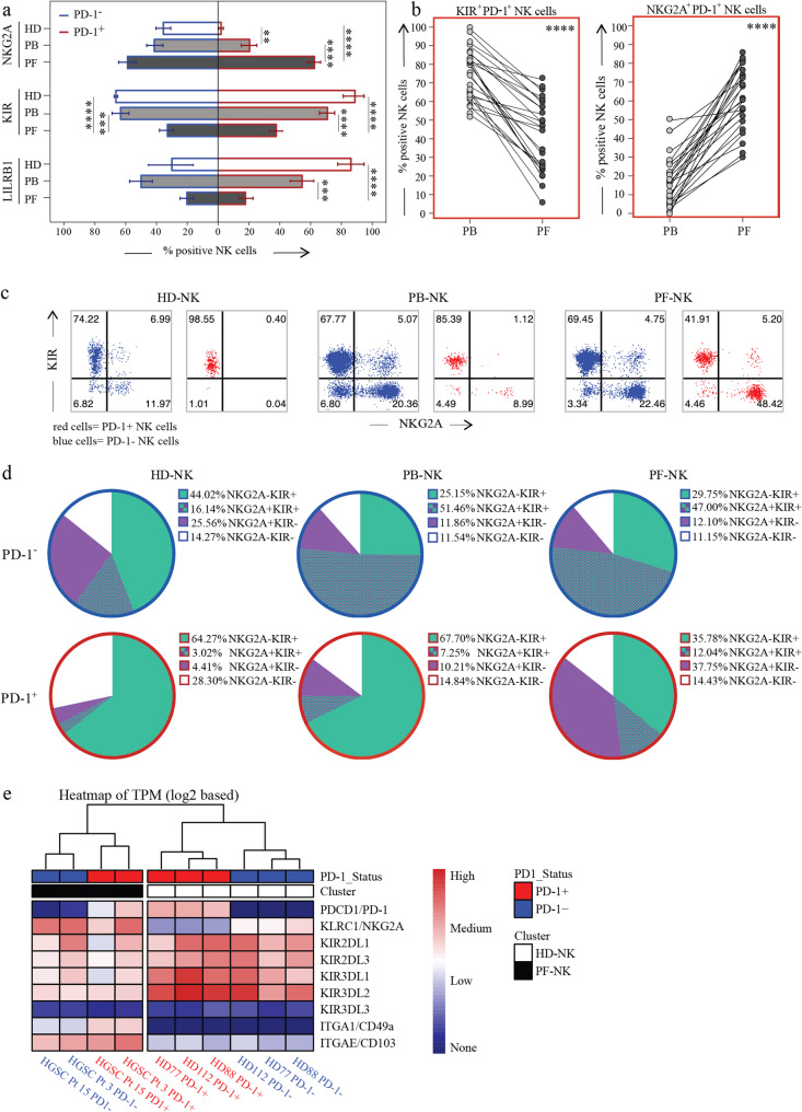
Results: We identified a distinct population of PD-1⁺ NK cells enriched in HGSC tumors and metastatic sites but absent in healthy donors. These cells, characterized by a CD56dimNKG2A⁺KIR⁺/⁻NKp46⁺CD57low phenotype, displayed impaired cytotoxicity against autologous HGSC targets, correlating with poorer prognosis. Crucially, this dysfunction was reversible upon combined blockade of PD-1/PD-L1, NKG2A, and KIRs. Spatial and molecular profiling revealed that these cells localize within PD-L1⁺/HLA-E⁺ tumor niches, suggesting that immune suppression is spatially and molecularly coordinated. Transcriptomic analysis confirmed their altered functional state and highlighted actionable checkpoint targets.

Conclusions: Our findings uncover a previously underappreciated population of dysfunctional PD-1⁺ NK cells in HGSC and demonstrate that their suppression is reversible through combinatorial checkpoint inhibition. These insights support the development of spatially-informed, NK-targeted immunotherapies for HGSC patients, particularly those resistant to T cell-based strategies.

Supplementary Information: The online version contains supplementary material available at 10.1186/s13046-025-03508-2.

Keywords: Immune checkpoint; Immunotherapy; Natural killer cells; Ovarian cancer; Programmed cell death 1 receptor; Tumor escape; Tumor infiltrating.


Conflict of interest statement

Declarations. Ethics approval and consent to participate: This study was carried out in accordance with the recommendations of the ethical standards of the institutional and/or national research committee. The protocol was approved by the ethics committee of the Liguria Region, Genova, Italy (n. 326/2018 and n127/2022-DB id12223 for OC patients and n. 39/2012 for healthy donors, by Spedali Civili of Brescia institutional ethical board (n. 1892/2010 for OC patients). All subjects gave written informed consent in accordance with the Declaration of Helsinki. Consent for publication: All authors have approved the submitted version. Competing interests: Eric Vivier and Pascale Andrè are employees of Innate Pharma, all other authors present no Conflicts of Interest.

Figures

Fig. 1
Fig. 1
PD-1 expression pattern in NK cells derived from HGSC patients and correlation with patient outcomes. a. Side-by-side expression of the PD-1 immune checkpoint (IC) on NK cells in the peripheral blood of healthy donors (HD-NK, white dots), peripheral blood (PB-NK, light gray dots) and peritoneal fluid (PF-NK, dark gray dots) of HGSC patients (HD n=200, HGSC patients n=60). b. Pie chart showing the ratio of subjects with a PD-1+ NK cell population greater than 1.5% in HD-NK, and in PB-NK and PF-NK of HGSC patients (HD n=200, HGSC patients n=60). c. Fold change of expression of analyzed ICs comparing the mean expression in HD the mean expression of PB and PF from HGSC patients (N>20). d. Expression of PD-1 on NK cells in the PB of a representative HD (HD-NK) and in the PB (PB-NK) and PF (PF-NK) of a representative HGSC patient. PD-1+ NK cells are in red. e. Percentage of seropositivity for HCMV in serum derived from HD (top) and HGSC patients (bottom) expressing PD-1 on NK cells (n=23). f. Different fluorescence intensity of PD-1 staining on PD-1+ cells of HD-NK and PF-NK g. Representation of Transcripts per Million (TPM) of PD-1 transcripts in PD-1+ and PD-1- NK cells of 3 HD and 2 PF samples. Gate strategy: CD45+CD3-CD19-CD14-CD56+(PD-1+/-) **: p< 0.01, ***: p<0.001, ****: p<0.0001
Fig. 2
Fig. 2
Correlation between PD-1 expression and patient survival.a. Correlation between PD-1 expression on NK cells derived from the PF of HGSC patients analyzed. Patient outcome was categorized in six stages (0-5), ranking from complete recovery to death within one year of diagnosis. b. Kaplan–Meier plots of progression-free survival in patients characterized by high (red) or low (blue) levels of PD-1 transcripts in bulk transcriptomic analysis of HGSC tissue. Data from HGSC patients for whom clinical and gene expression information was available (n=379), downloaded from cBioportal for Cancer. OS: overall survival
Fig. 3
Fig. 3
Co-expression of PD-1 with KIRs and NKG2A in HD and in PB and PF of HGSC patients. a. Expression of NKG2A, KIRs and LILRB1 on PD-1-CD56dim NK cells (blue outline) and PD-1+CD56dim NK cells (red outline) in HD-NK (white bars), and NK cells from PB (PB-NK, light gray bars), and PF (PF-NK, dark gray bars) of HGSC patients (n=25). b. Variation in the co-expression of KIRs (left) and NKG2A (right) with PD-1 between PB-NK (light gray dots) and PF-NK (dark gray dots) of HGSC patients. PB and PF samples from the same patient are connected by a black line (n=25). c, d. Representative dot plots (c) and pie charts (d) showing the distribution of KIRs and NKG2A in PD-1-NK cells (blue outline) and PD-1+NK cells (red outline) in HD-NK (left), PB-NK (center), and PF-NK (right) (n=25). e. Heatmap representing relative Transcripts per Million (TPM) of RNA transcripts coding for relevant NK cell markers on sorted PD-1+ (red label) and PD-1- (blue label) NK cells from three HD (white label) and two PF samples (black label). Gate strategy: panels a, c, d: CD45+CD3-CD19-CD14-CD56dimPD-1+/PD-1-; panel b: CD45+CD3-CD19-CD14-CD56dimPD-1+. **: p<0.01, ***: p<0.001, ****: p<0.0001
Fig. 4
Fig. 4
Peculiar PD-1+ NK subsets detectable in HSGOC patients. a. PD-1+ NK cells (red cells) in relation to CD57, NKG2A, KIRs and NKp46 surface expression in a representative HD (HD-NK, left), and a representative HGSC patient (PB-NK, center, and PF-NK, right). b Expression levels of the indicated markers (NKG2A, KIRs, NKp46 and CD57) on PD-1- (blue outline) and PD-1+ NK cell subsets (red outline) in HD (HD-NK, white bars) and HGSC patients (PB-NK, light gray bars and PF-NK, dark gray bars) (n=25). c. UMAP representation of the co-expression of important NK cell markers on NK cells from a representative HD (white area) and the PB (light gray) and PF (dark gray) of a representative HGSC patient, identifying four PD-1+ populations characterized by: I) a KIR+/-NKG2A-CD57+ phenotype characteristic of HD and PB; II) a KIR+NKG2A-CD57- phenotype characteristic of PF; III) a KIR+NKG2A+CD57- phenotype characteristic of PF; IV) a KIR-NKG2A+ CD57- phenotype characteristic of PF. Gate strategy: panel a, c: CD45+CD3-CD19-CD33-CD14-CD127-CD56dim; panel b: CD45+CD3-CD19-CD33-CD14-CD127-CD56dimPD-1+/-. **: p<0.01, ***: p<0.001, ****: p<0.0001
Fig. 5
Fig. 5
Proliferation assay of NK cells derived from the PF of HGSC patients. a. Proliferation evaluated with a CFSE dilution assay of PD-1- (blue column) and PD-1+ (red column) HD-NK (left) and PF-NK (right) (n=6). b. Proliferation evaluated with a CFSE dilution assay of PD-1- (blue column) and PD-1+ (red column) HD-NK (left) and PF-NK (right) stratified based on co-expression of the classical IC KIR and NKG2A (n=6). Gate strategy: CD45+CD3-CD19-CD14-CD56+
Fig. 6
Fig. 6
HGSC cells phenotype and ligand expression. a. Expression of inhibitory (HLA-I, HLA-E, PD-L1, PD-L2) and activating (B7-H6, Nectin-2, PVR) ligands on the surface of tumor cells from the PF and the primary tumor of HGSC patients (n=6). b. UMAP representation of most of the analyzed ligands on one representative PF and primary tumor. Gate strategy: 7AAD-CD45-CD14-CD90-CD140a-EPCAM+. c. (A-E) Multiplex immunohistochemical analysis of a representative primitive HGSC tissue showing extensive expression of HLA-E and PD-L1 on CK7+ cancer cells. (A’-E’) Enlargement of the area boxed in A showing that the tumor area is infiltrated by lymphocytes (arrows in A’). Scale bars are indicated in each panel
Fig. 7
Fig. 7
Proliferation assay of NK cells derived from the PF of HGSC patients. (a) Proliferation evaluated with a CFSE dilution assay of PD-1- (blue column) and PD-1+ (red column) HD-NK (left) and PF-NK (right) (n = 6). (b) Proliferation evaluated with a CFSE dilution assay of PD-1- (blue column) and PD-1+ (red column) HD-NK (left) and PF-NK (right) stratified based on co-expression of the classical IC KIR and NKG2A (n = 6). Gate strategy: CD45+CD3CD19CD14CD56+
Fig. 8
Fig. 8
PD-1+ NK cells infiltrate the tumor tissue and are more abundant in metastatic niche. a. Multiplex immunohistochemical analysis of a representative HGSC case showing infiltrating NK cells (NKp46+) expressing PD-1 and NKG2A and expression of PD-L1 and HLA-E on tumor cells (CK7+) in both ovarian and metastatic tissue. (A-H) Primary HGSOC showing co-expression of NKp46 (A,D), PD-1 (B,D), and NKG2A (C,D) on tumor infiltrating lymphocytes and co-expression of HLA-E (E,H), PD-L1 (F,H), and CK7 (G,H) on cancer cells. (A’-H’). Metastatic HGSOC showing co-expression of NKp46 (A’,D’), PD-1 (B’,D’), and NKG2A (C’,D’) on tumor infiltrating lymphocytes and co-expression of HLA-E (E’,H’), PD-L1 (F’,H’), and CK7 (G’,H’) on cancer cells. In all panels, the most prominent tumor cell mass is outlined. Arrows indicate NKp46+/PD-1+/NKG2A NK cells. Inserts in A–D and A’–D’ show cells indicated by yellow arrows at higher magnification. Scale bars are indicated in the Figure. b, c Expression of PD-1 on NK cells in the PB (PB-NK), PF (PF-NK), primary tumor (T-NK), and metastasis (MT-NK) of HGSC patients (n=7) and a representative HGSC patient. 0.0001 p: ****. Linear regression is shown. d, e. Representative dot plots (d) and pie charts (e) showing the distribution of KIRs and NKG2A in PD-1+NK cells in PB (PB-NK), primary tumor (T-NK), the PF (PF-NK), and metastasis (MT-NK) of HGSC patients (n=6). Gate strategy: panel b, c: CD45+CD3-CD19-CD14-CD56dim; panel d: CD45+CD3-CD19-CD14-CD56dim PD-1+

References

    1. Slatnik CLP, Duff E. Ovarian cancer: ensuring early diagnosis. Nurse Pract. 2015;40(9):47–54. - PubMed
    1. Momenimovahed Z, Tiznobaik A, Taheri S, Salehiniya H. Ovarian cancer in the world: epidemiology and risk factors. Int J Womens Health. 2019;11:287–99. - PMC - PubMed
    1. cancerresearchuk.org. https://www.cancerresearchuk.org/health-professional/cancer-statistics/s...
    1. Bowtell DD, Böhm S, Ahmed AA, Aspuria PJ, Bast RC, Beral V, et al. Rethinking ovarian cancer II: reducing mortality from high-grade serous ovarian cancer. Nat Rev Cancer. 2015;15(11):668–79. - PMC - PubMed
    1. Kuroki L, Guntupalli SR. Treatment of epithelial ovarian cancer. BMJ. 2020;371:m3773. - PubMed

LinkOut - more resources