

Use of MRI-double inversion recovery sequence for early diagnosis of multiple sclerosis: case series
- PMID: 40877867
- PMCID: PMC12395631
- DOI: 10.1186/s12883-025-04386-z
Use of MRI-double inversion recovery sequence for early diagnosis of multiple sclerosis: case series
Abstract
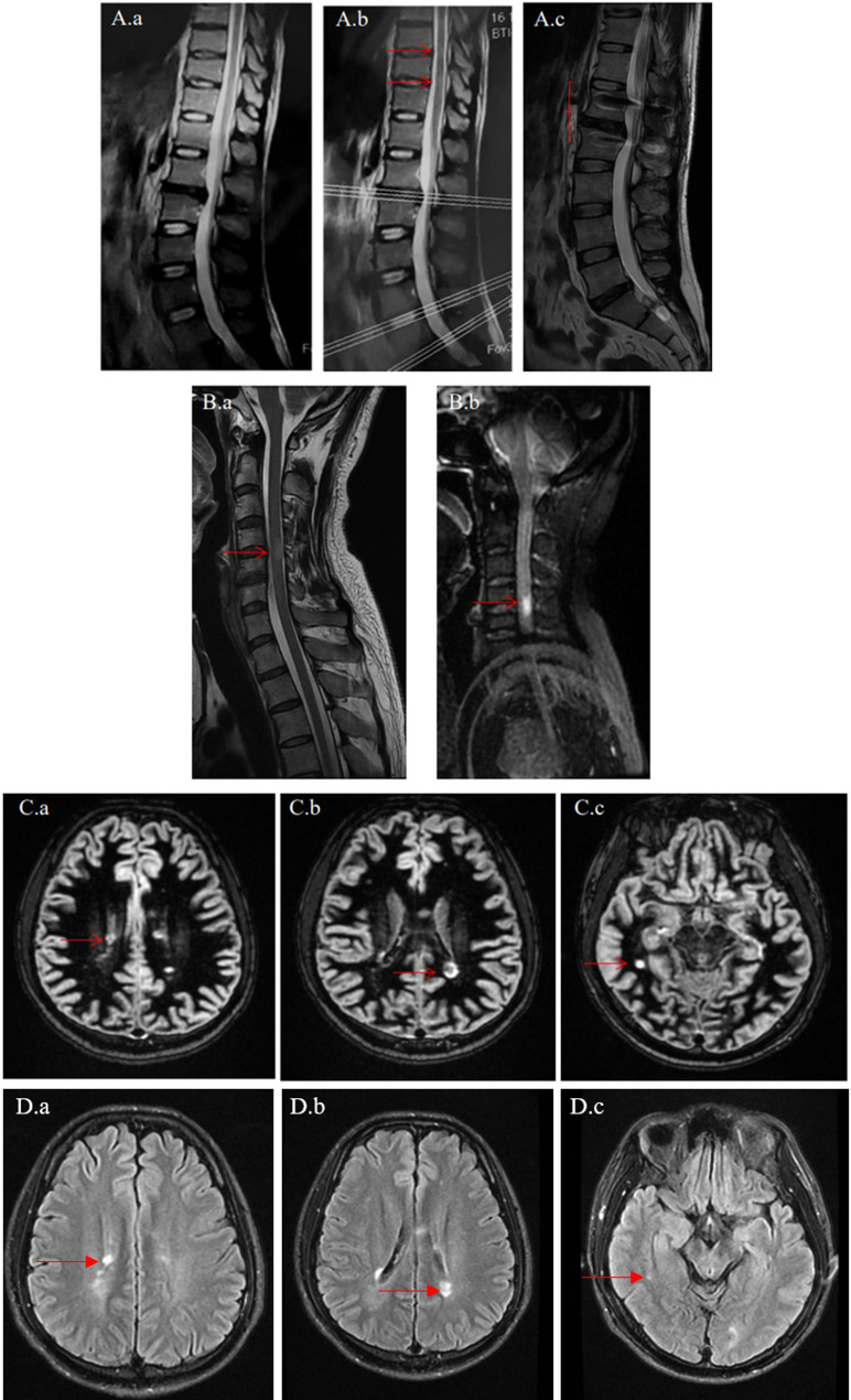
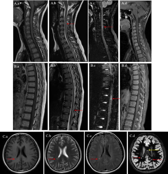
The choice of additional Double Inversion Recovery (DIR) Magnetic Resonance Imaging (MRI) sequences may affect the judgment of the presence and nature of lesions. Herein, we reported three cases initially presenting with non-typical multiple sclerosis (MS) symptoms, in which the final diagnosis of MS was confirmed after incorporating spinal cord and brain DIR MRI sequence. The sequence revealed more conspicuous lesions compared to conventional T2-weighted imaging or T2-weighted fluid-attenuated inversion recovery (T2WI/FLAIR) sequences. Consequently, the patients received an earlier definitive diagnosis and timely treatment. Our cases demonstrated the additional DIR sequences for the patients with non-typical MS symptoms were reasonable and may be helpful.
Keywords: Double inversion recovery sequence; Magnetic resonance imaging; Multiple sclerosis.
Conflict of interest statement
Declarations. Ethics approval and consent to participate: The study conformed to the Declaration of Helsinki. The research protocol was approved by the Institutional Review Committee of Shanghai East Hospital. The study was conducted in accordance with relevant code of ethics and regulations. All patients agreed to participate in this study. Each patient’s writing informed consent was obtained before examination. And all patients’ information will be kept confidential. Consent for publication: The participants have consented to the submission of the article to the journal. In addition, written informed consent for publication of clinical details and involved images was obtained from the three patients or their parents. Competing interests: The authors declare no competing interests.
Figures

Similar articles
-
Targeted magnetic resonance imaging (tMRI) of small changes in the T1 and spatial properties of normal or near normal appearing white and gray matter in disease of the brain using divided subtracted inversion recovery (dSIR) and divided reverse subtracted inversion recovery (drSIR) sequences.Quant Imaging Med Surg. 2023 Oct 1;13(10):7304-7337. doi: 10.21037/qims-23-232. Epub 2023 Aug 15. Quant Imaging Med Surg. 2023. PMID: 37869282 Free PMC article. Review.
-
Histopathology-validated recommendations for cortical lesion imaging in multiple sclerosis.Brain. 2020 Oct 1;143(10):2988-2997. doi: 10.1093/brain/awaa233. Brain. 2020. PMID: 32889535 Free PMC article.
-
Detection of multiple sclerosis lesions in the cervical cord: which of the MAGNIMS 'mandatory' non-gadolinium enhanced sagittal sequences is optimal at 3T?Neuroradiol J. 2021 Dec;34(6):600-606. doi: 10.1177/19714009211017787. Epub 2021 May 20. Neuroradiol J. 2021. PMID: 34014786 Free PMC article.
-
Comparison of Unenhanced and Gadolinium-Enhanced Imaging in Multiple Sclerosis: Is Contrast Needed for Routine Follow-Up MRI?AJNR Am J Neuroradiol. 2019 Sep;40(9):1476-1480. doi: 10.3174/ajnr.A6179. Epub 2019 Aug 22. AJNR Am J Neuroradiol. 2019. PMID: 31439627 Free PMC article.
-
The use of 7T MRI in multiple sclerosis: review and consensus statement from the North American Imaging in Multiple Sclerosis Cooperative.Brain Commun. 2024 Oct 9;6(5):fcae359. doi: 10.1093/braincomms/fcae359. eCollection 2024. Brain Commun. 2024. PMID: 39445084 Free PMC article. Review.
References
-
- Dobson R, Giovannoni G. Multiple sclerosis - a review. Eur J Neurol. 2019;26(1):27–40. 10.1111/ene.13819. - PubMed
-
- The Lancet Neurology. Multiple sclerosis under the spotlight. Lancet Neurol. 2021;20(7):497. 10.1016/S1474-4422(21)00170-8. - PubMed
-
- Patel S, Dimaandal I, Kuhns T, et al. Double inversion recovery to detect cervical spinal cord multiple sclerosis lesions. J Neuroimaging. 2023;33(4):521–6. 10.1111/jon.13100. - PubMed
Grants and funding
LinkOut - more resources
Full Text Sources
