. 2025 Aug 29;16(1):466.
doi: 10.1186/s13287-025-04604-y.

Cardioprotective effects of extracellular vesicles from hypoxia-preconditioned mesenchymal stromal cells in experimental pulmonary arterial hypertension

Affiliations

Cardioprotective effects of extracellular vesicles from hypoxia-preconditioned mesenchymal stromal cells in experimental pulmonary arterial hypertension

Renata Trabach Santos et al. Stem Cell Res Ther. .

Abstract

Background: During pulmonary arterial hypertension (PAH), cardiac cells develop a hypertrophic and apoptosis-resistant phenotype. Mesenchymal stromal cell (MSC) therapy has been shown to mitigate pulmonary vascular remodeling in PAH; however, successful application is limited by low potency and the need for a high number of MSCs. MSCs exposed to hypoxia release more extracellular vesicles (EV)s with different content than normoxia. We aimed to evaluate the proteomic profile and therapeutic effects of EVs derived from normoxia- and hypoxia-preconditioned MSCs on cardiac tissue remodeling in experimental PAH.

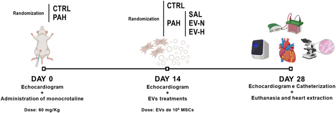
Methods: Isolated bone marrow MSCs were subjected to normoxia (N, 21%O2) or hypoxia (H, 1%O2) for 48 h and EVs were collected from the MSCs by ultracentrifugation. Proteomic data of the EVs were reanalyzed using PatternLab for Proteomics 5.0. Thirty-two male Wistar rats were randomly assigned to PAH plus intraperitoneal monocrotaline (60 mg/kg) or control (CTRL) with saline. On day 14, PAH animals received saline (1 mL/kg; PAH-SAL), EV-N (EVs from 1 × 10⁶ MSCs; PAH-EV-N) or EV-H (EVs from 1 × 10⁶ MSCs; PAH-EV-H) by jugular vein. On day 28, right ventricular systolic pressure (RVSP), pulmonary acceleration time/pulmonary ejection time (PAT/PET) ratio, right ventricle (RV) outflow diameter, and right ventricular hypertrophy (RVH) index were evaluated. The heart was harvested for histologic and molecular biology analyses.

Results: Among 695 proteins identified, 203 were present only in EV-H and 51 in EV-N. EV-H was enriched in proteins involved in the negative regulation of mitogen-activated protein kinase and apoptosis pathways. On day 28, both EV-N and EV-H therapies decreased RVSP compared with PAH-SAL (32 ± 5 mmHg and 29 ± 4 mmHg versus 39 ± 2 mmHg; p < 0.01). Only EV-H increased PAT/PET, reduced RV outflow diameter, and the RVH index compared with PAH-SAL. The expressions of c-Myc, a marker of myocardial injury, and p-GSK3β-Ser9, a proliferative marker, were higher in the PAH-SAL group than in the CTRL group. EV-N and EV-H decreased c-Myc expression, but only EV-H significantly reduced p-GSK3β-Ser9.

Conclusion: EV-N and EV-H reduced RVSP, but only EV-H improved RVH and RV outflow diameter, increased the PAT/PET ratio, and downregulated GSK3β protein levels. EVs from hypoxia-preconditioned MSCs demonstrated greater cardioprotective effects than those from normoxia-conditioned MSCs.

Keywords: Cardiac remodeling; Extracellular vesicles; Hypoxic pre-conditioning; Mass spectrometry; Mesenchymal stromal cells; Monocrotaline; Proteomic profile; Ventricle hypertrophy.


Conflict of interest statement

Declarations. Competing interest: The authors have no competing interests in the information described in this article.

Figures

Fig. 1
Fig. 1
Experimental protocol. CTRL, control; EV-H, extracellular vesicles from mesenchymal stromal cells subjected to hypoxia; EV-N, extracellular vesicles from mesenchymal stromal cells subjected to normoxia; EVs, extracellular vesicles; MCT, monocrotaline; MSCs, mesenchymal stromal cells; PAH, pulmonary arterial hypertension; SAL, saline. Monocrotaline dose, 60 mg/kg; extracellular vesicle dose, originating from 1 × 106 mesenchymal stromal cells
Fig. 2
Fig. 2
Echocardiographic representative images and functional parameters on day 28. (A) Upper row shows pulmonary artery Doppler and the lower row displays the diameter of the right ventricle outflow tract obtained in the long axis; (B) PAT/PET ratio; (C) RV outflow diameter measured via echocardiography on day 28 of the experimental protocol; and (D) right ventricular hypertrophy index calculated as the ratio of the right ventricular weight to the combined weight of the left ventricle and septum (RV/LV + S), adjusted for body weight. Yellow outline, peak blood flow in the pulmonary artery; yellow connector, right ventricle outflow diameter. CTRL, control; EV-H, extracellular vesicles from mesenchymal stromal cells subjected to hypoxia; EV-N, extracellular vesicles from mesenchymal stromal cells subjected to normoxia; PAH, pulmonary arterial hypertension; PAT, pulmonary acceleration time; PET, pulmonary ejection time; SAL, saline; n = 8 per group. Comparisons were done using one-way ANOVA followed by the Tukey multiple comparison test (p < 0.05); *significance versus CTRL, #significance versus PAH-SAL, &significance versus PAH-EV-N. Data are presented as means ± standard deviation
Fig. 3
Fig. 3
Right ventricular systolic pressure (RVSP) measurements on day 28. (A) Representative curves and (B) graph of RVSP measurements. CTRL, control; EV-H, extracellular vesicles from mesenchymal stromal cells subjected to hypoxia; EV-N, extracellular vesicles from mesenchymal stromal cells subjected to normoxia; PAH, pulmonary arterial hypertension; SAL, saline; n = 8 per group. Statistical comparisons were performed using one-way ANOVA followed by the Tukey multiple comparison test (p < 0.05); *significance versus CTRL, #significance versus PAH-SAL. Data are presented as means ± standard deviation
Fig. 4
Fig. 4
GSK3β inhibition and c-Myc expression on day 28. (A) Representative western blot analysis; (B) graph showing densitometric expression of phosphorylated GSK3β (p-GSK3β) at serine 9 (Ser9) relative to total GSK3β. GAPDH are also shown. (C) Real-time polymerase chain reaction analysis of c-Myc expression in cardiac tissue. Relative gene expression was calculated as the ratio of average gene expression levels compared with the reference gene 36B4 and expressed as fold change relative to the CTRL group. CTRL, control; EV-H, MSC-derived extracellular vesicles subjected to hypoxia; EV-N, MSC-derived extracellular vesicles subjected to normoxia; PAH, pulmonary arterial hypertension; SAL, saline; n = 5 per group. Statistical comparisons were performed using one-way ANOVA followed by the Tukey multiple comparison test (p < 0.05); *significance versus CTRL, #significance versus PAH-SAL; &significance versus PAH-EV-N. Data are presented as means ± standard deviation
Fig. 5
Fig. 5
Immune cell quantification in right ventricular sections on day 28. Representative photomicrographs and quantification of immune cells in right ventricle sections stained with hematoxylin-eosin at 400× magnification. Arrows indicate immune cells. CTRL, control; EV-H, MSC-derived extracellular vesicles subjected to hypoxia; EV-N, MSC-derived extracellular vesicles subjected to normoxia; PAH, pulmonary arterial hypertension; SAL, saline; n = 5 per group. Scale bar, 20 μm. Statistical comparisons were performed using one-way ANOVA followed by the Tukey multiple comparison test (p < 0.05); *significance versus CTRL, #significance versus PAH-SAL. Data are presented as means ± standard deviation
Fig. 6
Fig. 6
Macrophage infiltration in right ventricular sections on day 28. Representative photomicrographs and quantification of macrophages in right ventricle sections. (A) Total macrophage quantification; (B) phenotypic characterization of M1 macrophages; (C) phenotypic characterization of M2 macrophages. Immunohistochemistry was performed for CD68, iNOS, and mannose receptor (brown) at 400× magnification. Arrows indicate CD68, iNOS, and mannose receptor-positive cells. CTRL, control; EV-H, MSC-derived extracellular vesicles subjected to hypoxia; EV-N, MSC-derived extracellular vesicles subjected to normoxia; PAH, pulmonary arterial hypertension; SAL, saline; n = 5 per group. Scale bar, 20 μm. Statistical comparisons were performed using one-way ANOVA followed by the Tukey multiple comparison test (p < 0.05); *significance versus CTRL, #significance versus PAH-SAL. Data are presented as means ± standard deviation
Fig. 7
Fig. 7
Caspase-3-mediated programmed cell death in right ventricular sections on day 28. Representative photomicrographs and quantification of cleaved caspase-3 in right ventricle sections. Immunohistochemistry for cleaved caspase-3 (brown) at 400× magnification. Arrows indicate nuclear cleaved caspase-3 positive cells; arrowheads indicate cytoplasmic cleaved caspase-3 positive cells. CTRL, control; EV-H, MSC-derived extracellular vesicles subjected to hypoxia; EV-N, MSC-derived extracellular vesicles subjected to normoxia; PAH, pulmonary arterial hypertension; SAL, saline; n = 5 per group. Scale bar, 20 μm. Statistical comparisons were performed using one-way ANOVA followed by the Tukey multiple comparison test (p < 0.05); *significance versus CTRL, #significance versus PAH-SAL, &significance versus PAH-EV-N. Data are presented as means ± standard deviation

References

    1. Galiè N, McLaughlin VV, Rubin LJ, Simonneau G. An overview of the 6th world symposium on pulmonary hypertension. Eur Respir J. 2019;53(1):1802148. 10.1183/13993003.02148-2018 - PMC - PubMed
    1. Cao X, Fang X, Guo M, Li X, He Y, Xie M, et al. TRB3 mediates vascular remodeling by activating the MAPK signaling pathway in hypoxic pulmonary hypertension. Respir Res. 2021;22(1):312. 10.1186/s12931-021-01908-4 - PMC - PubMed
    1. Lv X, Li J, Wei R, Meng L, Kong X, Wei K, et al. Ethyl pyruvate alleviates pulmonary arterial hypertension via PI3K-Akt signaling. Mol Cell Biochem. 2025;480(2):1045–54. 10.1007/s11010-024-05020-1 - PubMed
    1. Gurbanov E, Shiliang X. The key role of apoptosis in the pathogenesis and treatment of pulmonary hypertension. Eur J Cardiothorac Surg. 2006;30(3):499–507. 10.1016/j.ejcts.2006.05.026 - PubMed
    1. Lafuse WP, Wozniak DJ, Rajaram MVS. Role of cardiac macrophages on cardiac inflammation, fibrosis and tissue repair. Cells. 2020;10(1):51. 10.3390/cells10010051 - PMC - PubMed

MeSH terms

LinkOut - more resources