. 2025 Aug 28;15(1):124.
doi: 10.1186/s13578-025-01457-z.

Enriched environment inhibits GLT-1 ubiquitination by downregulating SMURF1 to attenuate ischemic brain injury induced excitotoxicity

Affiliations

Enriched environment inhibits GLT-1 ubiquitination by downregulating SMURF1 to attenuate ischemic brain injury induced excitotoxicity

Yi Zhang et al. Cell Biosci. .

Abstract

Background: Excitotoxicity induced by glutamate contributes significantly to ischemic brain injury. The role of enriched environment (EE) in promoting neurological recovery post-stroke is well-established, yet its impact on excitotoxicity remains unclear. This study aimed to elucidate the mechanisms through which EE modulates excitotoxicity in ischemic brain injury using in vivo and in vitro experiments.

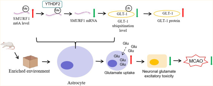
Results: EE exposure effectively reduced ischemic brain injury and glutamate-induced excitotoxicity in middle cerebral artery occlusion (MCAO) rat models. Notably, EE led to upregulated expression of GLT-1, which enhanced glutamate uptake by astrocytes and mitigated cytotoxicity and neuronal death induced by oxygen-glucose deprivation/reperfusion (OGD/R). Furthermore, EE suppressed the upregulation of SMURF1, which interacted with GLT-1 to facilitate its ubiquitin-mediated degradation and downregulate GLT-1 expression. Additionally, EE enhanced m6A modification of SMURF1, promoting YTHDF2-mediated mRNA decay and reducing SMURF1 expression. Knockdown of SMURF1 boosted GLT-1 expression, improving glutamate uptake and alleviating neuronal injury and death caused by OGD/R. Conversely, overexpression of SMURF1 or knockdown of GLT-1 attenuated the neuroprotective effects of EE against excitotoxicity in MCAO rats.

Conclusion: EE mitigates excitotoxicity in ischemic brain injury by inhibiting SMURF1 expression through m6A modification-YTHDF2-dependent pathways, thereby suppressing GLT-1 ubiquitin-mediated degradation and enhancing glutamate uptake.

Supplementary Information: The online version contains supplementary material available at 10.1186/s13578-025-01457-z.

Keywords: Enriched environment; Excitotoxicity; GLT-1; Ischemic brain injury; SMURF1.


Conflict of interest statement

Declarations. Ethics approval and consent to participate: All animal experiments were reviewed and approved by the Ethics Committee of the Nanchang University at Nanchang, China (Approval no. CDYFY-IACUC-202310QR019). Consent for publication: Not applicable. Conflict of interest: The authors declare no conflict of interest.

Figures

Fig. 1
Fig. 1
EE attenuates ischemic brain injury and excitotoxicity in rats byupregulating GLT-1 expression. MCAO rat models were established and housed on standardenvironment or EE. A, TTC staining to assess the infarct volume of rat brain tissues; B, mNSS to evaluate the neurological deficit; C, apoptosis of neurons detected by TUNELstaining; D, glutamate in the cerebrospinal fluid detected by ELISA; E, MAP2 and GLT-1expression detected by immunofluorescence; F, GLT-1 mRNA expression detected by qRT-PCR; G, protein expression of GLT-1, SYP and MAP2 detected by western blot.Comparison of panel A-G was determined by One-way analysis of variance, with Tukey’smultiple comparisons test for post hoc analysis. *, P< 0.05; **, P< 0.01; ***, P< 0.001.mNSS, modified Neurological Severity Scores; EE, enriched environment; MCAO, middlecerebral artery occlusion
Fig. 2
Fig. 2
Overexpression of GLT-1 increases glutamate uptake in astrocytes andinhibits OGD/R-induced cytotoxicity and neuron death. In vitro co-culture system of primary rat astrocytes and neurons were constructed for OGD/R and glutamate treatment.The astrocytes were infected with GLT-1 overexpressing lentivirus. A, qRT-PCR detectedthe transfection efficiency of GLT-1 overexpressing lentivirus; B, western blot detected thetransfection efficiency of GLT-1 overexpressing lentivirus; C, glutamate level in astrocytesdetected by ELISA; D, neuron viability assessed by CCK-8 assay; E, neurotoxicity detectedby LDH assay; F, neuron death detected by PI staining; G, protein expression of MAP2,SYP and caspase-3 detected by western blot. Comparison of panel A-B was analyzed usingStudent’s t-test whereas comparison of panel C-G was determined by One-way analysis ofvariance, with Tukey’s multiple comparisons test for post hoc analysis. *,P<0.05; **, P<0.01; ***, P「0.001. OGD/R, oxygen-glucose deprivation/reoxygenation; LDH, lactatedehydrogenase
Fig. 3
Fig. 3
SMURF1 inhibits GLT-1 expression by promoting its ubiquitination ubiquitinationdegradation, whereas EE downregulates SMURF1 expression. A, qRT-PCR to detect the level of SMURF1 mRNA; B: western blot to detect the protein level of SMURF1; C: Immunofluorescence to detect the co-localization of SMURF1 with GFAP; D: qRT-PCR todetect the level of SMURF1 mRNA; E: western blot to detect the level of SMURF1; F:western blot to detect the level of SMURF1 and GLT-1; G: western blot to detect the levelof GLT-1; H: western blot to detect the stability of GLT-1 protein after CHX treatment; I:Co-IP to detect the target binding of SMURF1 to GLT-1 protein; J: ubiquitination assay todetect the level of ubiquitination of GLT-1. Comparison of panel A-H was made using One-way analysis of variance test, with Tukey’s multiple comparisons test for post hoc analysis, *, P< 0.05; **; P< 0.01; ***, P< 0.001. EE, enriched environment
Fig. 4
Fig. 4
EE downregulates SMURF1 expression in astrocytes in an m6A-YTHDF2 dependent manner. A-B, SMURF1 m6A level detected by MeRIP; C-D, RIP verified the binding relationship between YTHDF2 and SMURF1 mRNA; E, qRT-PCR detected the YTHDF2 mRNA level; F, western blot detected the protein expression of YTHDF2; G-H, SMURF1 mRNA expression was detected after actinomycin D treatment; I, SMURF1mRNA expression was detected by qRT-PCR; J, western blot detected the protein expression of SMURF1. Comparison of panel B-F was analyzed by Student’s t-testwhereas comparison of panel A, G-J was determined by One-way analysis of variance,with Tukey’s multiple comparisons test for post hoc analysis. *, P< 0.05, **, P< 0.01, ***, P< 0.001
Fig. 5
Fig. 5
SMURF1 knockdown promotes the glutamate uptake in astrocytes viaupregulating GLT-1 expression to inhibit OGD/R induced toxicity and apoptosis. A,western blot detected SMURF1 and GLT-1 protein expression; B, ELISA detected theglutamate level in astrocytes; C, CCK-8 assay detected neuron activities; D, LDH assayassessed the toxicity; E, PI staining detected neuron death; F, western blot detected MAP2 SYP and caspase-3 protein expression. Comparison of panel A-F was analyzed One-wayanalysis of variance, with Tukey’s multiple comparisons test for post hoc analysis. *, P< 0.05, **, P< 0.01, ***, P< 0.001
Fig. 6
Fig. 6
EE suppresses ischemic nerve damage in rats through SMURF1/GLT-1axis.A, Cerebral infarction volume was assessed by TTC staining; B, nerve injury wasscored by mNSS; C, neurons was observed after Nissl staining; D, neuron apoptosis wasassessed by TUNEL staining; E, MAP2 expression was detected by immunofluorescence; F, protein expression of SYP and MAP2 was detected by western blot. Comparison of panelA-F was analyzed using One-way analysis of variance with Tukey’s multiple comparisonstest for post hoc analysis. *, P< 0.05, **, P< 0.01, ***, P< 0.001
Fig. 7
Fig. 7
EE attenuate glutamate excitotoxicity in MCAO rats throughSMURF1/GLT-1 axis. A, CSF was detected by ELISA; B, GLT-1 mRNA level was detectedby qRT-PCR; C, GLT-1 protein expression was detected by western blot; D, GLT-1 levelwas assessed by immunofluorescence. Comparison of panel A-D was analyzed using One-way analysis of variance with Tukey’s multiple comparisons test for post hoc analysis. *, P< 0.05, **, P< 0.01, ***, P< 0.001
Fig. 8
Fig. 8
Enriched environment reduces SMURF1 expression by increasing m6A level to decrease YTHDF2-mediated SMURF1 mRNA, while SMURF1 can promote the ubiquitinated degradation of GLT-1 and decrease GLT-1 expression. Knockdown of SMURF1 promotes glutamate uptake in astrocytes and inhibits cytotoxicity and neuron death by directly upregulating GLT-1 expression

Similar articles

References

    1. Sveinsson OA, Kjartansson O, Valdimarsson EM. Cerebral ischemia/infarction - epidemiology, causes and symptoms. Laeknabladid. 2014;100(5):271–9. - PubMed
    1. Dietz RM, Dingman AL, Herson PS. Cerebral ischemia in the developing brain. J Cereb Blood Flow Metab. 2022;42(10):1777–96. - PMC - PubMed
    1. Arnalich-Montiel A, Burgos-Santamaria A, Pazo-Sayos L, Quintana-Villamandos B. Comprehensive management of stroke: from mechanisms to therapeutic approaches. Int J Mol Sci. 2024;25(10). - PMC - PubMed
    1. Kannan A, Delgardo M, Pennington-FitzGerald W, Jiang EX, Christophe BR, Connolly ES Jr. Pharmacological management of cerebral ischemia in the elderly. Expert Opin Pharmacother. 2021;22(7):897–906. - PubMed
    1. Sakai S, Shichita T. Inflammation and neural repair after ischemic brain injury. Neurochem Int. 2019;130:104316. - PubMed

LinkOut - more resources