

Deep-Learning-Based Automated REM Sleep Detection in Patients With REM Sleep Behavior Disorder: Is It Reliable?
- PMID: 40878305
- PMCID: PMC12411298
- DOI: 10.3988/jcn.2025.0053
Deep-Learning-Based Automated REM Sleep Detection in Patients With REM Sleep Behavior Disorder: Is It Reliable?
Abstract
Background and purpose: Rapid eye movement (REM) sleep without atonia makes it difficult to detect REM sleep stages using electromyography in patients with REM sleep behavior disorder (RBD). The objectives of this study were to propose an automated REM sleep detector that requires only electroencephalography (EEG) and electrooculography (EOG) data, and to evaluate its performance using real-world polysomnography (PSG) data in RBD patients.
Methods: This multicenter study used 310 PSG datasets obtained from 5 tertiary hospitals. The data were divided into RBD (n=200) and non-RBD (n=110), as well as, into Parkinson's disease (PD) with RBD (n=76), PD without RBD (n=46), idiopathic RBD (iRBD) (n=124), and healthy controls (n=64). An automated computerized REM detection algorithm was implemented using U-Sleep's publicly available pretrained network.
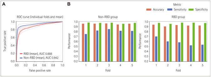
Results: The U-Sleep-based REM sleep-detection algorithm correctly identified REM sleep with an area under the receiver operating characteristic curve (AUC) of 0.90±0.14. The classification performance of the REM sleep detector differed significantly between RBD and non-RBD patients (AUC=0.88±0.13 vs. 0.93±0.14, p=0.007). The REM sleep detector accurately classified REM sleep in the order of healthy controls, PD without RBD, iRBD, and PD with RBD, with AUC values of 0.94±0.02, 0.92±0.03, 0.90±0.02, and 0.86±0.02, respectively.
Conclusions: Our U-Sleep-based REM sleep detector based on only EEG and EOG data showed good performance in detecting REM sleep. However, it performed considerably worse in RBD, especially in PD with RBD. Using transfer learning with fine-tuning by expert review, a high-performance REM sleep-detecting system will be realized.
Keywords: REM sleep behavior disorder; REM sleep detector; REM sleep without atonia; automated algorithm.
Copyright © 2025 Korean Neurological Association.
Conflict of interest statement
The authors have no potential conflicts of interest to disclose.
Figures


References
-
- Sateia MJ. International classification of sleep disorders-third edition: highlights and modifications. Chest. 2014;146:1387–1394. - PubMed
-
- Schenck CH, Mahowald MW. REM sleep behavior disorder: clinical, developmental, and neuroscience perspectives 16 years after its formal identification in SLEEP. Sleep. 2002;25:120–138. - PubMed
-
- Olson EJ, Boeve BF, Silber MH. Rapid eye movement sleep behaviour disorder: demographic, clinical and laboratory findings in 93 cases. Brain. 2000;123:331–339. - PubMed
-
- St Louis EK, Boeve AR, Boeve BF. REM sleep behavior disorder in Parkinson’s disease and other synucleinopathies. Mov Disord. 2017;32:645–658. - PubMed
Grants and funding
LinkOut - more resources
Full Text Sources
