. 2025 Oct;80(10):2710-2725.
doi: 10.1111/all.16672. Epub 2025 Aug 29.

Molecular Signatures and Functional Pathways of Human Monocytes and Macrophages in Allergy: An EAACI AllergoOncology Scoping Review

Affiliations

Molecular Signatures and Functional Pathways of Human Monocytes and Macrophages in Allergy: An EAACI AllergoOncology Scoping Review

Rodolfo Bianchini et al. Allergy. 2025 Oct.

Abstract

AllergoOncology explores the intersection of allergic diseases and cancer, focusing on shared immune mechanisms. While monocytes and macrophages are extensively studied in cancer, their roles in allergic diseases remain underexplored. To address this gap, we conducted a scoping review to systematically characterize the molecular landscape and related pathways of human monocytes and macrophages in allergy. An automated search of PubMed and Web of Science databases retrieved 4668 unique articles, which were manually curated based on predefined inclusion and exclusion criteria, yielding 138 eligible studies. From these, we identified 451 molecules associated with monocyte and macrophage responses across allergic disorders. Data analyses revealed a research bias towards blood-derived monocytes, underrepresentation of tissue-resident macrophages, and limited inclusion of miRNAs. Semantic similarity and pathway enrichment analyses highlighted a common molecular signature across major allergic disorders, with consistent enrichment in interleukin signaling and immune activation pathways. To enhance reproducibility and translational utility for researchers and clinicians, we developed ALO•HA, a web application for interactive data exploration. This overview of monocyte and macrophage molecular responses in human allergy underscores the need for integrative, human-focused approaches to better define their roles, and to guide future therapeutic strategies in allergic diseases and at the interface with oncology.

Keywords: AllergoOncology; allergy; data‐driven bioinformatics; macrophages; monocytes.


Conflict of interest statement

All authors have read and approved the manuscript. Any potential conflicts of interest related to the manuscript are listed here: S.N.K. is the founder and shareholder of Epsilogen Ltd. (formerly IGEM Therapeutics Ltd.) and has received funds from IGEM Therapeutics Ltd/Epsilogen Ltd. H.J.B., and M.G. are employed through a fund provided by Epsilogen Ltd. J.C. has been employed through a fund provided by Epsilogen Ltd. S.N.K. and H.J.B. are inventors of patents on antibody technologies. S.N.K. reports grants from Worldwide Cancer Research (24‐0087); Guy's and St Thomas's Foundation Trust Charity Melanoma Special Fund (573); the British Skin Foundation (006/R/22); the Biotechnology and Biological Sciences Research Council (BB/T008709/1); Cancer Research UK (C30122/A11527; C30122/A15774); the Cancer Research UK King's Health Partners Centre at King's College London (C604/A25135); the CRUK City of London Centre Award (C7893/A29290); Breast Cancer Now (147; KCL‐BCN‐Q3); the Medical Research Council (MR/L023091/1; MR/R015643/1; MR/V049445/1); Innovate UK (51746); S.N.K. has projects supported by the King's Health Partners Centre for Translational Medicine. The views expressed are those of the author(s) and not necessarily those of King's Health Partners. O.P. received research grants from MINECO, Ministerio de Ciencia e Innovación, CAM, Inmunotek S.L., Novartis, and AstraZeneca and fees for giving scientific lectures or participation in Advisory Boards from: AstraZeneca, Pfizer, GlaxoSmithKline, Inmunotek S.L., Novartis, Sanofi‐Genzyme and Regeneron. E.J.J. is inventor in EP2894478; “LCN2 as a tool for allergy diagnostic therapy”, EP 14150965.3, Year: 01/2014; US 14/204,570, immunoBON, owned by Biomedical International R+D GmbH, Vienna, Austria, of which E.J.J. is shareholder.

Figures

FIGURE 1
FIGURE 1
Ontogeny of monocytes and macrophages during embryogenesis and postnatal life. The three overlapping developmental waves of monocytes and macrophages in the human embryo are illustrated, referenced to Carnegie stages (CS) and days post‐conception (dpc), together with the postnatal bone marrow pathway. In the first wave (1, orange lines): CS7‐CS10, ~16–22 dpc), yolk sac (YS) hemogenic endothelium generates primitive macrophage progenitors that migrate directly into tissues (e.g., brain), differentiating into microglia and extra‐embryonic macrophages such as Hofbauer cells, prior to the emergence of hematopoietic stem cells (HSCs). The second wave (2, red lines): CS11‐CS23, ~23–56 dpc) arises from erythro‐myeloid progenitors (EMPs) in the YS at CS11 (~23 dpc), which enter the circulation, colonize the fetal liver at CS12‐CS13 (~28–30 dpc), and differentiate into fetal monocytes. These monocytes seed tissues (lungs, skin, liver), establishing long‐lived, tissue‐resident macrophage populations. The third wave (3, blue lines): CS14‐CS17, ~30–42 dpc onward) originates from hemogenic endothelium in the aorta‐gonad‐mesonephros (AGM), generating HSCs that seed the fetal liver and, later, the bone marrow, producing fetal monocytes and contributing postnatally to monocyte/macrophage pools. In postnatal life (gray line), bone marrow–derived HSCs generate circulating monocyte subsets: Classical (CD14++CD16), intermediate (CD14++CD16+), and non‐classical (CD14+CD16++). Each of them with distinct roles in immune surveillance and inflammation. Classical monocytes (CCR2+) are recruited to inflamed tissues via the CCR2/CCL2 axis, where they differentiate into short‐lived monocyte‐derived macrophages or dendritic cells (key points: Long‐lived, fetal‐origin macrophages seed tissues prenatally, short‐lived, postnatal‐origin macrophages derive from circulating monocytes during inflammation. Figure created with BioRender.
FIGURE 2
FIGURE 2
The human macrophage polarization spectrum and functional diversity. (A) In vitro polarization spectrum of monocyte‐derived macrophages (M0), in two main phenotypic extremes: M1‐like (pro‐inflammatory) and M2‐like (anti‐inflammatory and tissue repair). M1‐like macrophages are activated by stimuli such as IFN‐γ, lipopolysaccharide (LPS), or microbial products, leading to the production of cytokines (e.g., IL‐12, TNFα, IL‐1, IL‐6, and IL‐23), and chemokines (e.g., CXCL9 and CXCL10), along with the expression of surface markers such as CD80/86, MHCII, and CCR7, reflecting their role in immune activation and pathogen elimination. In contrast, M2‐like macrophages display functional diversity across distinct subtypes: M2a, induced by IL‐4 or IL‐13, is characterized by high CD206/CD209 expression, and are CD23hi and FcεRIhi, mediating tissue repair and anti‐inflammatory responses. M2b, driven by immune complex stimulation with TLR ligands or IL‐1R agonists, produces IL‐10, TNFα, and IL‐6 and expresses CD86 and CD206dim, contributing to immunoregulation. M2c, stimulated by IL‐10 and TGFβ, express markers like CD163, MerTK, TLR7, and TLR8, contributing to tissue remodeling and resolution of inflammation. M2d, induced by exposure to adenosine or hypoxia‐inducible factors (HIFs), is linked to angiogenesis and immunosuppression, with intermediate expression of FcεRI (FcεRIint) and CD23 (CD23int), alongside IL‐10 and IL‐6 production. The figure highlights the dynamic functional plasticity of macrophages in response to distinct microenvironmental signals, shaping their roles in immunity, inflammation, and tissue homeostasis. (B) Representation of the continuum of macrophage activation states. Monocytes differentiate into macrophages displaying a remarkable plasticity, as they transition along a dynamic continuum of activation states rather than fitting into the rigid M1/M2 dichotomies. Figure created with BioRender.
FIGURE 3
FIGURE 3
Schematic representation outlining the literature search, screening, and selection processes. (A) Systematic pipeline used in this study. (B) Inclusion and exclusion criteria applied during the literature search and full‐text review.
FIGURE 4
FIGURE 4
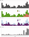
Trends in allergy research covering publications, study approaches, cell types, and identified molecules. (A) Annual publication trends highlighting the number of articles investigating monocyte and/or macrophage features in allergic disorders. (B) Proportions of studies employing targeted vs. untargeted methods reveal the predominant use of targeted approaches. (C) Distribution of studies by cell type, categorizing them as focusing on monocytes, macrophages, or both. (D) Annual trends in the number of molecules studied showed a substantial increase in molecular investigations in recent years, particularly with the adoption of high‐throughput methodologies. The star next to 2024 indicates that only studies published until July of that year were included in the search.
FIGURE 5
FIGURE 5
Associations between allergic disorders and sample sources. Sankey diagram shows the relationships between allergic disorders (left) and their corresponding sample sources (right). The thickness of each link and the associated percentages indicate the frequency of specific disorders and sample source combinations within the 138 selected articles. Disorder abbreviations and corresponding MeSH terms in parentheses: Allergic Alveolitis (AAlveolitis, D000542), Allergic Asthma (AAsthma, D001249), Allergic Bronchopulmonary Aspergillosis (ABA, D001229), Allergic Contact Dermatitis (ACD, D017449), Atopic Dermatitis (AD, D003876), Allergic Rhinitis (AR, D065631), Food Allergy (FA, D005512), and Immediate Hypersensitivity (IH, D006969).
FIGURE 6
FIGURE 6
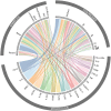
Interconnections among the most studied molecules, allergic disorders, and sample sources. Chord diagram visualizing the associations between top 20 molecules selected based on their occurrence across the analyzed articles, along with allergic disorders and sample sources. Molecules are sorted within the plot according to their total number of associations with disorders and sample sources, ranked from highest to lowest. The width of the links represents the frequency of co‐occurrence, while gray sections adjacent to molecules, disorders and samples summarize the aggregated connections, highlighting their significance in allergy research. Disorder abbreviations: AAlveolitis, Allergic Alveolitis; AAsthma, Allergic Asthma; ABA, Allergic Bronchopulmonary Aspergillosis; ACD, Allergic Contact Dermatitis; AD, Atopic Dermatitis; AR, Allergic Rhinitis; FA, Food Allergy; and IH, Immediate Hypersensitivity.
FIGURE 7
FIGURE 7
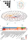
Shared and unique molecular and functional features in monocyte and macrophage responses across the most represented allergic disorders. (A) Disease Ontology semantic similarity matrix showing similarity scores among Allergic Asthma (AAsthma), Allergic Rhinitis (AR), Atopic Dermatitis (AD), Food Allergy (FA) and Allergic Alveolitis (AAlveolitis). (B) Venn diagram visualizing the number of shared and unique molecules among the 5 main disorders. (C) Network illustrating molecules associated with multiple disorders, where nodes represent molecules and disorders; edges indicate molecule‐disorders associations, and node size reflects the degree of interconnectivity. Colored nodes and edges correspond to specific allergic conditions. (D) Heatmap of Reactome enrichment analysis showing enriched pathways across disorders. Gray bars on the top represent the total number of unique genes per enriched pathway. Gray bars on the right show the total number of unique genes present in each disorder. The top bar annotation categorizes pathways into 5 top‐level Reactome pathways.
FIGURE 8
FIGURE 8
Overview of the scoping review workflow and outcomes. The graphical summary illustrates the process from literature curation and annotation to molecular and immunological insights, highlighting key annotations, associations, and pathway analyses, as well as access to the interactive ALO•HA platform. [Correction added on 16 September 2025, after first online publication: A new figure 8 and caption have been added.]

References

    1. Jensen‐Jarolim E., Bax H. J., Bianchini R., et al., “AllergoOncology—The Impact of Allergy in Oncology: EAACI Position Paper,” Allergy 72, no. 6 (2017): 866–887. - PMC - PubMed
    1. Jensen‐Jarolim E., Bax H. J., Bianchini R., et al., “AllergoOncology: Opposite Outcomes of Immune Tolerance in Allergy and Cancer,” Allergy 73, no. 2 (2018): 328–340. - PMC - PubMed
    1. Bergmann C., Poli A., Agache I., et al., “AllergoOncology: Danger Signals in Allergology and Oncology: A European Academy of Allergy and Clinical Immunology (EAACI) Position Paper,” Allergy 77, no. 9 (2022): 2594–2617. - PMC - PubMed
    1. Turner M. C., Radzikowska U., Ferastraoaru D. E., et al., “AllergoOncology: Biomarkers and Refined Classification for Research in the Allergy and Glioma Nexus‐A Joint EAACI‐EANO Position Paper,” Allergy 79, no. 6 (2024): 1419–1439. - PubMed
    1. Sreejit G., Fleetwood A. J., Murphy A. J., and Nagareddy P. R., “Origins and Diversity of Macrophages in Health and Disease,” Clinical & Translational Immunology 9, no. 12 (2020): e1222. - PMC - PubMed

Publication types