Thyroblastoma in Pregnancy: Expanding the Cytomorphological Spectrum of a Novel DICER1 -Associated Entity, a Case Report and Literature Review
- PMID: 40879299
- PMCID: PMC12402541
- DOI: 10.1002/dc.25504
Thyroblastoma in Pregnancy: Expanding the Cytomorphological Spectrum of a Novel DICER1 -Associated Entity, a Case Report and Literature Review
Abstract
Introduction: Thyroblastoma is a rare, aggressive thyroid neoplasm newly classified in the 2022 WHO Classification of Endocrine Tumors. It is characterized by embryonal, multilineage morphology and DICER1 mutations. Fewer than 15 well-characterized cases have been reported, with limited cytological descriptions.
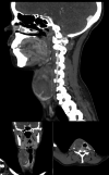
Case presentation: A 19-year-old pregnant woman presented with a rapidly enlarging right thyroid mass. Fine needle aspiration biopsy (FNAB) revealed a highly cellular, triphasic aspirate composed of primitive epithelium arranged in macrofollicular structures, spindled mesenchymal cells within a myxoid matrix, and small round blastemal cells. Immunocytochemistry showed TTF-1 and PAX8 positivity in epithelial cells, synaptophysin and SALL4 in blastemal cells, and myogenin in both spindled and blastemal components, supporting a cytological diagnosis of thyroblastoma. She underwent total thyroidectomy and neck dissection during the second trimester. Histology confirmed thyroblastoma with extensive lymph node metastases. Chemotherapy was initiated during pregnancy but discontinued due to neutropenic complications. She delivered a healthy infant and remained disease-free at her 12-month follow-up.
Molecular findings: Next-generation sequencing of tumor DNA revealed two somatic DICER1 mutations: a known hotspot missense mutation, c.5437G>C; p.E1813Q, and a novel splice-site variant, c.734+1G>T, predicted to disrupt normal splicing and protein function. No pathogenic variants were found in germline DNA, supporting a somatic origin.
Conclusion: This case expands the cytological and molecular spectrum of thyroblastoma and highlights the value of FNAB in early recognition. Awareness of this rare entity and its diagnostic features is essential to avoid misclassification and ensure timely management. The novel DICER1 splice-site mutation further contributes to the evolving molecular landscape of thyroblastoma.
Keywords: DICER1; Bethesda thyroid cytopathology; fine needle aspiration biopsy; malignant thyroid teratoma; molecular cytopathology; thyroblastoma.
© 2025 The Author(s). Diagnostic Cytopathology published by Wiley Periodicals LLC.
Conflict of interest statement
The authors declare no conflicts of interest.
Figures


References
-
- Agaimy A. N. V., Sobrinho‐Simoes M. T., and WHO Classification of Tumours Editorial Board , “Thyroid Tumours,” in WHO Classification of Endocrine and Neuroendocrine Tumours, vol. 10, 5th ed., ed. Tallini G. (International Agency for Research on Cancer, 2022).
-
- Rabinowits G., Barletta J., Sholl L. M., Reche E., Lorch J., and Goguen L., “Successful Management of a Patient With Malignant Thyroid Teratoma,” Thyroid 27, no. 1 (2017): 125–128. - PubMed
-
- Rooper L. M., Bynum J. P., Miller K. P., et al., “Recurrent DICER1 Hotspot Mutations in Malignant Thyroid Gland Teratomas: Molecular Characterization and Proposal for a Separate Classification,” American Journal of Surgical Pathology 44, no. 6 (2020): 826–833. - PubMed
-
- Agaimy A., Witkowski L., Stoehr R., et al., “Malignant Teratoid Tumor of the Thyroid Gland: An Aggressive Primitive Multiphenotypic Malignancy Showing Organotypical Elements and Frequent DICER1 Alterations‐Is the Term “Thyroblastoma” More Appropriate?,” Virchows Archiv 477, no. 6 (2020): 787–798. - PMC - PubMed
Publication types
MeSH terms
Substances
LinkOut - more resources
Full Text Sources
Medical

