. 2025 Oct 8;16(10):e0151625.
doi: 10.1128/mbio.01516-25. Epub 2025 Aug 29.

PTP1B deficiency in myeloid cells increases susceptibility to Candida albicans systemic infection by modulating antifungal immunity

Affiliations

PTP1B deficiency in myeloid cells increases susceptibility to Candida albicans systemic infection by modulating antifungal immunity

Bethany Allen et al. mBio. .

Abstract

Invasive candidiasis, primarily caused by Candida albicans, poses a significant threat to immunocompromised patients, with high mortality rates. Understanding how immune responses to Candida albicans are mounted and controlled is fundamental to developing new therapeutic strategies. Protein tyrosine phosphatase 1B (PTP1B) is a regulator of immunoreceptor signaling and downstream inflammatory and metabolic responses and a pharmaceutical target. Here, we reveal a critical role for myeloid cell-intrinsic PTP1B in antifungal immunity. Mice lacking PTP1B in myeloid cells (LysM PTP1B-/-) were significantly more susceptible to systemic C. albicans infection, exhibiting lower survival, greater weight loss, and elevated fungal burdens in the kidney, liver, and brain. These mice also showed heightened proinflammatory mRNA expression in organs and increased kidney tubular inflammation, with increased leukocyte infiltration and chemokine production, contributing to immunopathology. Neutrophils from LysM PTP1B-/- mice displayed impaired maturation in infected kidneys and reduced reactive oxygen species production in vitro. Proteomic profiling of infected bone marrow-derived macrophages (BMDMs) revealed significant enrichment of type I interferon-regulated proteins in the absence of PTP1B. These BMDMs showed impaired phagocytosis, reduced killing capacity, and lower viability during infection, phenotypes recapitulated in human macrophages treated with a pharmacological PTP1B inhibitor. Collectively, our findings highlight PTP1B as a key modulator of innate immune responses to C. albicans, balancing antifungal activity and systemic toxicity with inflammation and metabolic fitness. Boosting specific PTP1B-dependent pathways may offer new strategies for enhancing host defense while minimizing fungal-induced immunopathology.IMPORTANCESystemic Candida albicans infections are a leading cause of hospital-acquired morbidity and mortality, particularly in immunocompromised individuals and patients receiving immunosuppressive treatments. Despite antifungal therapies, outcomes remain poor, underscoring the need to better understand host factors that control fungal clearance. Protein tyrosine phosphatase 1B (PTP1B) is a key intracellular regulator of immune and metabolic signaling. This study identifies a critical role for myeloid PTP1B in antifungal defense and susceptibility to systemic C. albicans infection. Loss of PTP1B impairs neutrophil and macrophage function, disrupts inflammatory balance, and compromises pathogen clearance. These findings reveal PTP1B as a central modulator of immune responses to C. albicans and highlight its potential as a target for host-directed therapies to improve outcomes in systemic fungal infections.

Keywords: Candida albicans; infections; macrophages; protein tyrosine phosphatase 1B.


Conflict of interest statement

The authors declare no conflict of interest.

Figures

Fig 1
Fig 1
Myeloid cell PTP1B regulates antifungal immunity in vivo. (A) Survival curve of WT and LysM PTP1B−/− mice after C. albicans challenge. WT and LysM PTP1B−/− mice (nine mice per group) were infected intravenously with 2 × 10⁵ CFU of C. albicans, and percentage survival was calculated over the time period indicated. Data were pooled from two independent experiments. *P < 0.05, Mantel-Cox log-rank test. (B) WT and LysM PTP1B−/− mice were infected with 2 × 10⁵ CFU of C. albicans and sacrificed 3 days post-infection; WT (n = 9) and LysM PTP1B−/− (n = 8). Data are pooled from two independent experiments for each time point. Body weights were recorded for each mouse and expressed as percentage weight relative to total body weight prior to infection; one-way analysis of variance with Tukey multiple comparison test; *P < 0.05. (C–H) Organs were harvested, and the number of fungal colony-forming units per gram of tissue (CFU/g) was calculated for kidney, brain, and liver, either 24 h (C–E) or 3 days (F–H) post-infection; *P < 0.05, **P < 0.01 by Mann-Whitney U-test.
Fig 2
Fig 2
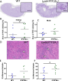
LysM PTP1B−/− mice exhibit greater fungal lesions and inflammation than WT mice. (A) Kidney sections were stained with PAS to visualize C. albicans hyphae, and examples are shown for whole sections and magnified fungal lesions (scale bar shows 100 µm). The area of PAS staining, 3 days post-infection for kidney (B) and brain (C), as determined by ImageJ analysis, was calculated as a percentage of the total area of the section. *P < 0.01, Mann-Whitney U-test. (D) hematoxylin and eosin stain (H&E) stained 3 days post-infection kidney section from WT and PTP1B−/− mice showing inflammation as indicated by white arrows. The percentage of inflamed tubules was calculated for WT and LysM PTP1B−/− in H&E-stained kidney sections from mice culled 24 h (E) and 3 days (F) post-infection; *P < 0.05, by Student’s t-test with Welch’s correction.
Fig 3
Fig 3
LysM PTP1B−/− mice show increased tissue and systemic cytokine expression. WT and LysM PTP1B−/− mice were infected with 2 × 10⁵ CFU of C. albicans and sacrificed 24 h post-infection (A–D) in two separate experiments (n = 3 in each experiment) or 3 days post-infection (E–H) in two separate experiments (n = 3, n = 6 for WT; n = 3, n = 5 for LysM PTP1B−/−). Fold change (2-ΔΔCt) in expression of Tnf, Il6, Il1b, and Il10 mRNA in LysM PTP1B−/− in kidney (A, E), liver (B, F), brain (C, G), and spleen (D, H) was calculated relative to median expression in WT. Bars represent mean ± SEM; *P < 0.05 represents significant difference between LysM PTP1B−/− and WT. Serum was collected from mice 24 h post-infection, and levels of cytokines were analyzed by multiplex arrays. Serum levels of cytokines are expressed as relative levels from mean fluorescence intensity values minus background for TNF-alpha (I), IL-6 (J), and IL-10 (K). Values from individual experiments and mean are shown.
Fig 4
Fig 4
LysM PTP1B−/− mice show increased chemokine expression and infiltrating immune cells in the infected kidney and peritoneum. Expression levels of chemokines cxcl1, cxcl2, and ccl2, as determined by real-time RT-qPCR, in WT and LysM PTP1B−/− mice kidneys isolated 24 h post-infection with C. albicans (A). Results were normalized to the expression of the housekeeping genes and are presented as fold change relative to median expression in WT. *P < 0.05, Student's t-test. Correlations between proportion of CD45+ leukocytes out of total viable cells and fungal burdens (CFU/g) in kidney of WT (blue) and LysM PTP1B−/− (green) mice at 24 h post systemic infection with Candida albicans (B). Pearson correlation and linear regression were used for statistical analysis. WT and LysM PTP1B−/− mice were infected systemically with 2 × 10⁵ CFU of C. albicans and sacrificed 24 h post-infection in two separate experiments (n = 3 in each experiment). Flow cytometry was used to analyze the expression of immune cell markers in the kidney (C–F) and spleen (G–I). Total immune cells were those viable cells that were CD45+. Inflammatory monocytes were defined as CD45+ CD11b+ Ly6 Chi (D, G), and neutrophils were defined as CD45+ CD11b+ Ly6G+ (E, H). Each cell population was expressed as a percentage of viable CD45+ leukocytes in the organ. CD45+ CD11b+ Ly6G+ neutrophils were analyzed for levels of Ly6G expression, shown by geometric mean fluorescence intensity (MFI) of PE-Ly6G antibody (F, I). *P < 0.05. WT and LysM PTP1B−/− mice were infected intraperitoneally with C. albicans (1 × 10⁶ CFU) and peritoneal cells isolated 4 h post-infection and analyzed by flow cytometry. The total number of isolated peritoneal cells (J), CD45+ cells (K), inflammatory monocytes defined by CD45+ CD11b+ and Ly6Chi (L), and neutrophils as defined by CD45+ CD11b+ and Ly6Ghi (M). *P < 0.05. Mann-Whitney U-test.
Fig 5
Fig 5
PTP1B controls the antifungal activity of neutrophils. Bone marrow-derived neutrophils isolated from age- and sex-matched LysM PTP1B−/− and WT mice were infected with live serum-opsonized (A, C, E, G) or non-opsonized (B, D, F, H) C. albicans at a multiplicity of infection (MOI) of 10. Cells from two to three separate mice were pooled for each experiment (n = 5-6 separate experiments performed on separate days). Bone marrow-derived neutrophils were incubated with C. albicans for 5 or 30 min, and the phagocytic activity was measured by flow cytometry to determine the percentage fungal uptake (A, B) and the phagocytic index, determined as the geometric mean fluorescence intensity (MFI) (C, D). Isolated bone marrow-derived neutrophils were infected with live opsonized (E) or unopsonized (F) C. albicans at an MOI of 10 for 5 or 30 min, and viability was assessed using flow cytometry. Cells were gated for Ly6G (PE) expression, and cells negative for eFluor 780 (APC-Cy7) were defined as live. For A–F, paired lines represent the average values from LysM PTP1B−/− and WT neutrophils from individual experiments. To determine killing efficiency, bone marrow-derived neutrophils were infected with live C. albicans yeast cells (MOI = 0.5) for 1 h and lysed. C. albicans at the same concentration, but without cells, was incubated for the same time as a control. The lysates were plated, and viable C. albicans cells were scored by counting the colonies formed on the plates (CFU) 24 h later (G, H). The percentage of killing = (colony number of control − colony number of neutrophil-treated)/colony number of control × 100. Bone marrow-derived neutrophils were infected with live C. albicans yeast cells (MOI = 10), and kinetics of ROS production was measured via a luminescence-based assay. Luminescence was measured as relative luminescence units (RLUs) to empty plate wells over 2 h (example shown in panel I, n = 1, mean ± SEM of quintuplicate technical replicates). Raw peak RLUs (J) and peak RLUs relative to WT (K) were recorded for each experiment (n = 7, mean ± SEM). *P < 0.05, two-way analysis of variance (A–F); Student’s t-test (G, H, K).
Fig 6
Fig 6
PTP1B regulates antifungal effector functions of macrophages. (A) BMDMs from age- and sex-matched mice were infected with live C. albicans yeast cells (multiplicity of infection (MOI) = 0.5) for 3 h, then lysed. Fungal CFUs were calculated per milliliter of lysis solution (n = 5) and used to define killing efficacy. BMDMs were infected with live fluorescein isothiocyanate-labeled C. albicans yeast cells (MOI = 1) for 3 h, and percentage uptake (B) and phagocytic index (C) of C. albicans were measured via microscopy (n = 5). Cytokine levels in supernatants of C. albicans-stimulated bone marrow-derived macrophages for TNF-alpha (D), IL-10 (E), and IL-6 (F) exposed to either heat-killed C. albicans or zymosan (100 µg/mL), as determined by Enzyme-linked immunosorbent assay (ELISA). Data are represented as mean ± SD (n = 12 individual BMDM preparations). Significant differences in levels of cytokine between WT and PTP1B−/− BMDMs are shown; *P < 0.05, Student’s t-test.
Fig 7
Fig 7
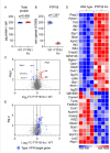
Deletion of PTP1B alters the proteome of C. albicans-stimulated macrophages. BMDMs from WT and LysM PTP1B−/− mice were infected with live C. albicans (multiplicity of infection (MOI) = 4) for 8 h. Cells were then lysed and the proteomes analyzed using data-independent acquisition (DIA)-based mass spectrometry as described in Materials and Methods. Separate cultures from four mice per genotype were analyzed. (A) Total protein content (pg/cell) was estimated for the copy number data derived from the mass spectrometry results; P-value was determined by a two-tailed Student's t-test. (B) Estimated copy numbers for Ptpn1 from the wild-type and LysM PTP1B−/− macrophages. (C) Volcano plot showing −log2 fold change in mean protein copy numbers per cell between C. albicans-infected WT and PTP1B−/− BMDMs where cut-off thresholds are plotted as vertical lines. Upregulated (red) and downregulated (blue) proteins were defined as having a fold change more than two standard deviations away from the median and a P-value of less than 0.01 with cutoff threshold shown as horizontal line. (D) Heat map for the z-scores of the proteins classed as upregulated or downregulated in panel C. (E) Volcano plot showing the regulation of potential type I interferon-regulated proteins, highlighted in blue, differentially expressed between the wild-type and LysM PTP1B−/− macrophages infected with C. albicans. Type I interferon-regulated proteins were defined based on a published transcription study in BMDMs (25).
Fig 8
Fig 8
Deletion of PTP1B alters the metabolic profile of BMDM that consequently decreases their viability and increases susceptibility to infection. (A) Blood glucose levels in uninfected and 24 h post C. albicans-infected (2 × 10⁵ yeast cells/mouse) WT and LysM PTP1B−/− mice. (B) Quantitative PCR analysis of metabolic gene expression in kidneys of 24 h post C. albicans-infected WT or LysM PTP1B−/− mice for glycolytic enzyme Pfkfb3, glucose importers SLC2A1 (GLUT1) and SLC5A2 (SGLT2), and gluconeogenic genes G6PC and PCK1. Results were normalized to the expression of the housekeeping genes and are presented as fold change relative to median value of WT BMDM. (C) A Cell Mito Stress Test was performed using a Seahorse XFe24 Analyzer and oxygen consumption rates (OCRs) measured with representative example of WT and LysM PTP1B−/− BMDM with and without heat-killed C. albicans activation shown. (D) Basal OCR, (E) ATP production by mitochondria, (F) spare respiratory capacity, (G) basal ECAR, and (H) maximum glycolytic capacity of macrophages were measured with or without heat-killed C. albicans in WT and LysM PTP1B−/− BMDM and normalized with cell numbers. Data are shown as mean ± SEM, n = 4–5 separate BMDM preparations. (I) Cell death as a percentage of total cells in WT and PTP1B−/− BMDM infected with live C. albicans MOI 1:1. (J) Cell death as a percentage of total human monocyte-derived macrophage following treatment with 0, 0.7, 1.5, or 3.0 µM PTP1B inhibitor MSI1436. (K) Cell death as a percentage of total cells in WT and PTP1B−/− BMDM infected without or with live C. albicans multiplicity of infection (MOI) 1:3 with or without addition of glucose 20 mM. Shown are mean values and SEM for five to six separate BMDM preparations, or three to four individual human monocyte preparations from each group performed in triplicate. * = P < 0.05, ** = P < 0.01; analysis of variance (ANOVA) (A, B, I) or ANOVA with Tukey’s multiple comparisons post hoc test (DH, J, K). For graph K, uninfected WT and LysM PTP1B−/− BMDM were significantly different from WT and LysM PTP1B−/− BMDM without and with glucose; however, significant differences are not marked for clarity.

References

    1. Denning DW. 2024. Global incidence and mortality of severe fungal disease. Lancet Infect Dis 24:e428–e438. doi: 10.1016/S1473-3099(23)00692-8 - DOI - PubMed
    1. Lass-Flörl C, Kanj SS, Govender NP, Thompson GR 3rd, Ostrosky-Zeichner L, Govrins MA. 2024. Invasive candidiasis. Nat Rev Dis Primers 10:20. doi: 10.1038/s41572-024-00503-3 - DOI - PubMed
    1. Brown GD, Denning DW, Gow NAR, Levitz SM, Netea MG, White TC. 2012. Hidden killers: human fungal infections. Sci Transl Med 4:165rv13. doi: 10.1126/scitranslmed.3004404 - DOI - PubMed
    1. Pathakumari B, Liang G, Liu W. 2020. Immune defence to invasive fungal infections: a comprehensive review. Biomed Pharmacother 130:110550. doi: 10.1016/j.biopha.2020.110550 - DOI - PubMed
    1. Kozicky LK, Sly LM. 2015. Phosphatase regulation of macrophage activation. Semin Immunol 27:276–285. doi: 10.1016/j.smim.2015.07.001 - DOI - PubMed

MeSH terms

Substances

Supplementary concepts

LinkOut - more resources