. 2025 Sep 2;122(35):e2506257122.
doi: 10.1073/pnas.2506257122. Epub 2025 Aug 29.

TIGER: A tdTomato in vivo genome-editing reporter mouse for investigating precision-editor delivery approaches

Affiliations

TIGER: A tdTomato in vivo genome-editing reporter mouse for investigating precision-editor delivery approaches

Samuel W Du et al. Proc Natl Acad Sci U S A. .

Abstract

In vivo genome editing has the potential to address many inherited and environmental disorders. However, a major hurdle for the clinical translation of genome editing is safe, efficient delivery to disease-relevant tissues. A modality-agnostic reporter animal model that facilitates rapid, precise, and quantifiable assessment of functional delivery and editing could greatly enhance the evaluation and translation of delivery technologies. Here, we present the development of the tdTomato in vivo genome-editing reporter (TIGER) mouse, a reporter strain that harbors an integrated and constitutively expressed mutated tdTomato gene in the Polr2a locus. The mutations (Q115X, Q357X) abolish fluorescence, but successful adenine base editing (ABE) or prime editing (PE) restores tdTomato fluorescence. This mouse model facilitates the tissue- and cell type-specific assessment of genome editing agent delivery. We describe several editing strategies validated in vitro and demonstrate efficient ABE and PE in vivo using viral and nonviral delivery vectors targeting four cell types within the mouse eye: the retinal pigment epithelium, photoreceptors, Müller glia, and the trabecular meshwork. We show direct editing characterization in the ocular tissues via in vivo and ex vivo two-photon confocal microscopy and verify the spectral and fluorescence lifetime properties of tdTomato reporter in other mouse tissues. Additionally, we demonstrate successful adeno-associated virus (AAV)-mediated PE of extraocular tissues, including hepatocytes, skeletal muscle, and brain neurons by intravenous injection. Thus, the TIGER mouse facilitates the direct development, comparison, and optimization of delivery platforms for efficient and productive ABE or PE broadly applicable in vivo across multiple tissues tested in this study.

Keywords: CRISPR; base editing; genome editing; prime editing; reporter.


Conflict of interest statement

Competing interests statement:K.P. is a consultant for Polgenix Inc. and serves on the Scientific Advisory Board at Hyperion Eye Ltd. D.R.L. is a consultant, co-founder and/or equity owner of Beam Therapeutics, Prime Medicine, Pairwise Plants, and nChroma Bio, companies that use or deliver genome or epigenome editing agents. The Broad Institute has filed patent applications on base and prime editing. The remaining authors declare no competing interests.

Figures
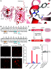
Fig. 1.
Fig. 1.
Validation of tdTomato editing strategies. (A) Left, AlphaFold2-predicted structure of tdTomato (23). The Gln115 and Gln357 residues, whose codons are replaced by nonsense TAG mutations, are represented in stick format on the ribbon diagram of the protein. Right, the Inset of ancestral DsRed chromophore structure determined by X-ray crystallography (24). (B) Genomic sequence of tdTomato. Upper sequence, mutant base in red; lower sequence, wild-type tdTomato. The PAM sequences for ABE (orange) and PE (blue) are highlighted. (C) Schematic diagram of TIGER construct stably expressed in HEK293T cells via lentiviral transduction. (D) Microscopy of TIGER HEK293T cells transfected with ABE or PE with cognate guide RNAs. (E) Amplicon sequencing of TIGER alleles by NGS, showing quantification of editing results. Mean ± SD. (F) Anti-α-tubulin and anti-RFP immunoblotting of HEK AAVS1 TIGER cell extracts obtained from cultures: 1) nontreated, 2) ABE RNP treated, 3) ABE eVLP treated. Predicted molecular weights of tdTomato: Nonedited, 13.1 kDa; N-terminal edited, 40.2 kDa; full-length, 54.2 kDa.
Fig. 2.
Fig. 2.
Design and construction of the TIGER mouse. (A) Strategy for knock-in of the mouse TIGER construct into the Polr2a locus. PCAG3.0, chicken beta-actin hybrid promoter; WPRE, woodchuck hepatitis virus posttranscriptional regulatory element; bGH, bovine growth hormone polyadenylation signal; Neo, neomycin resistance cassette. (B) TIGER genotyping gel. (C) Verification of absence of the rd1 mutation. (D) Verification of absence of the rd8 mutation. (E) Heatmap of Polr2a expression in mouse tissues by single-cell RNA sequencing [ln(CPM) of FACS data] from the Tabula Muris dataset (30).
Fig. 3.
Fig. 3.
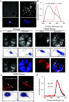
Two-photon characterization of tdTomato fluorescence properties in vivo. (A) Two-photon microscopy of HEK293T TIGER cells untreated or treated with ABE8e-N108Q eVLP. Top, fluorescence intensity signal; Bottom, phasor-plot FLIM analysis. The red circle in the phasor plots indicates the expected location of the tdTomato-FLIM signal. (B) Normalized emission spectrum of untreated (black) and eVLP-treated (red) cells. (C) Two-photon fundus imaging of anesthetized, live heterozygous TIGER mice either untreated (Left) or injected intravenously (Right) with dual-AAV2/8 PEmax. Upper panels, fluorescence intensity; Middle panels, phasor plot FLIM analysis; Bottom panels, FLIM in pseudocolor assigned by phasor plot analysis. (D) Phasor FLIM of different retinal cell types in the intact eyes of TIGER mice injected intravenously with dual-AAV2/8 PEmax. Upper panels, pseudocolor in FLIM (scale bar represents 50 µm); Lower panels, assignments in phasor plot analysis. (E) Normalized emission spectra of cells in the outer nuclear layer (ONL, black), and the RPE (red) in the eyes of PEmax-treated TIGER mice.
Fig. 4.
Fig. 4.
In vivo editing within the retinas of TIGER mice. (A) RPE flatmounts from the eyes of WT and TIGER mice treated subretinally with ABE-eVLP. (Scale bar represents 500 µm.) N = 14 for treated TIGER mice. (B) Microscopy of eye cryosection from an ABE-eVLP-treated TIGER mouse. (Scale bar represents 75 µm.) N = 14 for treated TIGER mice. (C) Two-photon 3D volumetric reconstruction of intact eye from a TIGER mouse treated with subretinal dual-AAV2/8 (scales provided in µm). RPE, retinal pigment epithelium; PR, photoreceptor; MG, Müller glia. N = 19 for treated TIGER mice. (D) Two-photon 3D volumetric reconstruction of the intact RPE of a TIGER mouse treated with subretinal ABE-eVLP (scales provided in µm). N = 14 for treated TIGER mice. (E) Microscopy of an eye cryosection from a TIGER mouse treated with subretinal dual-AAV2/8 PEmax. (Scale represents 500 µm.) The Inset is a magnified region of the cryosection. (Scale bar represents 75 µm.) ONH, optic nerve head. N = 19 for treated TIGER mice. (F) NGS amplicon sequencing analyzing precise correction and indel rates in four heterozygous TIGER eyes treated with dual-AAV2/8 PEmax (Left) and eight heterozygous TIGER eyes treated with ENVLPE+ PE VLPs (Right). (G) Two-photon 3D volumetric reconstruction of the intact RPE of a heterozygous TIGER mouse treated with subretinal ENVLPE+-PEmax-VLP (scales provided in µm). N = 8 for treated TIGER mice. (scales provided in µm). (H) Quantification via ImageJ analysis of maximum intensity projections of tdTomato+ cells per area in heterozygous TIGER mice treated with dual-AAV2/8 (blue, N = 5) or ENVLPE VLPs (purple, N = 3). RPE, retinal pigment epithelium; PR, photoreceptors; MG, Müller glia.
Fig. 5.
Fig. 5.
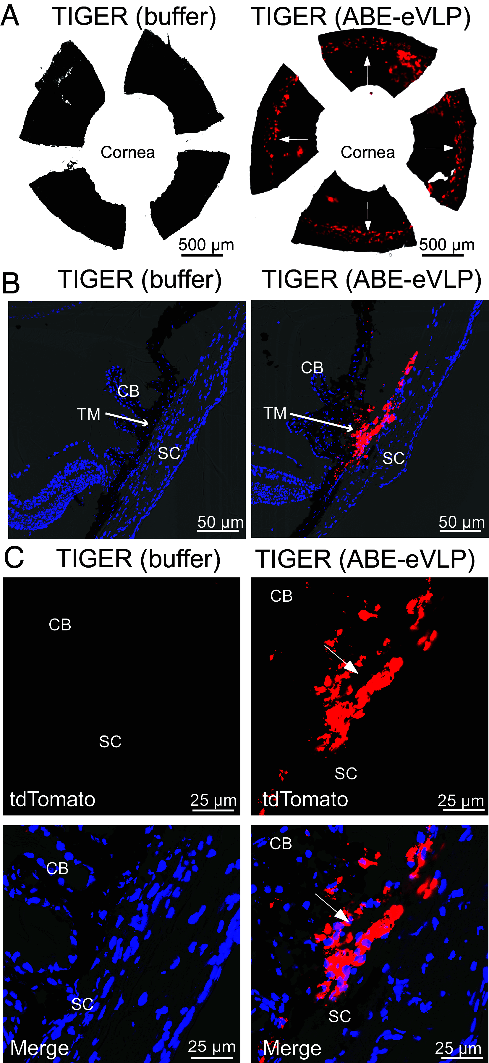
In vivo editing of the anterior segment from TIGER mice. (A) Anterior segment flatmount from a TIGER mouse injected intracamerally with ABE-eVLP- or buffer-treated control. White arrows point to the trabecular meshwork. (Scale bar represents 500 µm.) N = 3 for treated TIGER mice. (B) Low-magnification confocal imaging of cryosections of the anterior segment from a TIGER mouse treated with buffer, or injected intracamerally with ABE-eVLP. The white arrow points to the trabecular meshwork. CB, ciliary body; SC, Schlemm’s canal; TM, trabecular meshwork. (Scale bar represents 50 µm.) N = 7 for treated TIGER mice. DAPI, blue; tdTomato, red. (C) Higher magnification images of the sections shown in B. (Scale bar represents 25 µm.) DAPI, blue; tdTomato, red.
Fig. 6.
Fig. 6.
In vivo editing within the liver of TIGER mice. (A) Liver cryosections from TIGER mice injected intravenously with PBS (Left) or dual-AAV8 PEmax (Right), imaged for tdTomato. The Insets show the corresponding DAPI-counterstained sections. (Scale bar represents 500 µm.) N = 5 for treated TIGER mice. (B) High magnification liver cryosections from TIGER mice injected intravenously with dual-AAV8 PEmax. (Scale bar represents 50 µm.)
Fig. 7.
Fig. 7.
In vivo editing within the skeletal muscle of TIGER mice. Heterozygous TIGER mice were injected locally with 1 × 10¹¹ VG of dual AAV8 PEmax into the right gastrocnemius muscle. Three weeks after injection, the treated-right and untreated-left gastrocnemius muscles were collected, sectioned, and imaged for tdTomato editing and fluorescent rescue. (Scale bar represents 100 µm.)
Fig. 8.
Fig. 8.
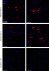
In vivo editing within the CNS of TIGER mice. (AD) Images show tdTomato expression in the olfactory bulb of a TIGER mouse following injection of AAV (A, B, and D) or PBS (C); (B) shows neuronal layers labeled with NeuN (green) and DAPI (purple), relative to the tdTomato as shown in (A). The most prominent labeling following dual-AAV8 PEmax injection was in the glomeruli (dotted outline) and resident neurons including mitral cells and their processes (arrow); the Inset shows a mitral cell layer neuron. (EN) Fields containing tdTomato expression in TIGER mice: (E) Anterior cingulate layer II; (F) cerebellar granule cell layer, with occasional cells in the Purkinje cell layer (Inset); (G) indusium griseum neuron; (H) axons in the cochlear nucleus; (I) axons in the deep inferior colliculus; (JL) neuronal perikarya in the trigeminal motor (J), facial motor (K), and mesenchephalic trigeminal nuclei (L) (Inset, higher magnification); arrows indicate cells double-labeled for NeuN. (M) tdTomato-expressing mossy fibers in the interposed deep cerebellar nucleus (arrowheads); and (N) tdTomato-expressing climbing fibers extending into the cerebellar molecular layer. The pattern is typical for these types of axons which encase Purkinje cell dendrites. Scale bars: panel (C) = 100 µm for (AC); panel (D) = 100 µm (50 µm for Inset); panels (E and F) = 40 µm (50 µm for Inset); panels (G and I) = 55 µm; panel (H) = 40 µm; panel (J) = 45 µm; panel (K) = 65 µm; panel (L) = 60 µm (45 µm for Inset); panel (M) = 15 µm; panel (N) = 45 µm. N = 5 for treated TIGER mice.

References

    1. Komor A. C., Kim Y. B., Packer M. S., Zuris J. A., Liu D. R., Programmable editing of a target base in genomic DNA without double-stranded DNA cleavage. Nature 533, 420–424 (2016). - PMC - PubMed
    1. Gaudelli N. M., et al. , Programmable base editing of A*T to G*C in genomic DNA without DNA cleavage. Nature 551, 464–471 (2017). - PMC - PubMed
    1. Anzalone A. V., et al. , Search-and-replace genome editing without double-strand breaks or donor DNA. Nature 576, 149–157 (2019). - PMC - PubMed
    1. Newby G. A., Liu D. R., In vivo somatic cell base editing and prime editing. Mol. Ther. 29, 3107–3124 (2021). - PMC - PubMed
    1. Raguram A., Banskota S., Liu D. R., Therapeutic in vivo delivery of gene editing agents. Cell 185, 2806–2827 (2022). - PMC - PubMed

LinkOut - more resources