Case Reports
. 2025 Oct 6;13(28):107759.
doi: 10.12998/wjcc.v13.i28.107759.

Magnetic resonance imaging spectrum of acute hemorrhagic leukoencephalitis: Four case reports

Affiliations
Case Reports

Magnetic resonance imaging spectrum of acute hemorrhagic leukoencephalitis: Four case reports

Ankit Shukla et al. World J Clin Cases. .

Abstract

Background: Acute hemorrhagic leukoencephalitis (AHLE), also known as Weston-Hurst syndrome, is a very rare and fulminant form of demyelinating disorder. It is considered a hyperacute and severe variant of acute disseminated encephalomyelitis. Clinically, patients present with fever, headache, seizures, and altered sensorium, which can rapidly progress to coma or death. Magnetic resonance imaging (MRI) is the investigation of choice and plays a pivotal role in diagnosing AHLE. The purpose of this article is to make readers familiar with the typical MRI features of AHLE and to discuss differentials.

Case summary: This case series reports the clinical presentation and typical neuroimaging findings in four patients diagnosed with AHLE. All patients presented with acute neurological symptoms, such as severe headaches, seizures, and altered consciousness, often following a history of fever suggesting an infectious etiology. Additionally, laboratory investigations demonstrated elevated levels of serum inflammatory markers and neutrophilic pleocytosis on cerebrospinal fluid analysis, supporting a post-infectious etiology. MRI findings consistently revealed characteristic white matter lesions with hemorrhagic foci and vasogenic edema, indicative of widespread demyelination characteristic of AHLE. The outcomes varied, with two patients surviving but experiencing neurological sequelae, while two others unfortunately succumbed to the disease. The clinical data, laboratory results, and imaging findings from this case series were systematically compared with those from previously published studies. The key similarities and differences in clinical presentation, imaging characteristics, and outcomes are presented in a tabulated format.

Conclusion: AHLE is associated with high morbidity and mortality rates, emphasizing the need for early recognition, prompt intervention, and multidisciplinary management. Further research is needed to explain the pathophysiological mechanisms underlying AHLE, identify potential biomarkers for early diagnosis, and develop targeted therapies to improve patient outcomes.

Keywords: Case report; Central nervous system; Demyelinating disease; Hemorrhage; Hurst’s disease; Leukoencephalitis; Magnetic resonance imaging.


Conflict of interest statement

Conflict-of-interest statement: All the authors report no relevant conflicts of interest for this article.

Figures
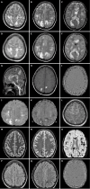
Figure 1
Figure 1
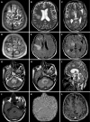
Magnetic resonance imaging brain findings in a 19-year-old female at initial presentation and on follow-up. A-C: Axial T2 weighted images showing multifocal hyperintensities in subcortical and deep white matter with corpus callosum involvement; D-F: Axial Fluid attenuated inversion recovery (FLAIR) images demonstrating confluent hyperintensities in bilateral cerebral white matter and corpus callosum; G: Sagittal T2-weighted image showing fluffy white matter and callosal hyperintensities; H and I: Axial T1 weighted image (H) showing internal hyperintense areas within white matter lesions, which on Susceptibility weighted imaging (SWI) phase image (I) are showing negative phase effect suggestive of hemorrhages; J and K: DWI and apparent diffusion coefficient images showing patchy diffusion restriction; L: Post-contrast T1-weighted image showing no enhancement; M and N: Follow-up T2-weighted images showing resolution of white matter lesions; O: Follow up SWI image showing multiple hemorrhagic residues in white matter; P and Q: Follow-up FLAIR images showing resolution of white matter lesions; R: Follow-up SWI phase image showing multiple residual hemorrhagic foci.
Figure 2
Figure 2
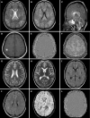
Magnetic resonance imaging brain findings in a 40-year-old male patient who presented with fever and altered sensorium. A-C: T2-weighted axial images showing multifocal subcortical and deep white matter hyperintensities in bilateral cerebral hemispheres associated with involvement of right hypothalamus; D-F: Corresponding axial Fluid attenuated inversion recovery (FLAIR) images showing similar findings as seen on T2-weighted images; G and H: Axial T2-weighted and FLAIR images showing hyperintensity involving pons and bilateral middle cerebellar peduncles; I: T2 weighted sagittal image showing hyperintense lesions involving midbrain, pons, medulla and scanned part of the cervical spinal cord; J: T1-weighted axial image showing hypointensity in the involved areas with internal hyperintense areas suggestive of hemorrhages; K: Susceptibility weighted imaging (SWI) axial phase image showing a negative phase effect within white matter lesions indicative of hemorrhages; L: T1-weighted post-contrast axial image showing no enhancement in the affected regions.
Figure 3
Figure 3
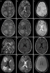
Magnetic resonance imaging brain findings of an 18-year-old female patient during hospitalization and on follow up. A: Axial T2 weighted image showing multifocal subcortical and deep white matter hyperintensities in the bilateral cerebral hemispheres; B: Axial Fluid attenuated inversion recovery (FLAIR) image showing multiple white matter hyperintensities; C: Sagittal T2 weighted image revealing involvement of the corpus callosum; D: Axial T1 weighted image showing hypointense lesions with internal, hyperintense foci in the affected areas; E: Axial Susceptibility weighted imaging (SWI) phase image demonstrating a negative phase effect, suggestive of hemorrhagic components. F: Axial post contrast T1 weighted image showing no obvious enhancement; G–J: Follow-up T2-weighted (G, H) and FLAIR (I, J) images showing resolution of previously noted white matter hyperintensities; K and L: Follow up SWI images revealing an increased number of hemorrhagic residues compared to initial imaging.
Figure 4
Figure 4
Magnetic resonance imaging brain findings in a 45-year old female patient. A and B: Axial T2-weighted images showing fluffy hyperintense lesions in subcortical and deep white matter; C: Axial Gradient echo sequence (GRE) image revealing blooming areas within white matter lesions, suggestive of hemorrhages; D and E: Axial fluid attenuated inversion recovery images showing multifocal hyperintense white matter lesions; F: Axial GRE image demonstrating hemorrhagic foci within the lesions; G: Sagittal T2-weighted image illustrating white matter hyperintensities; H and I: Axial T1-weighted image showing multifocal hypointense lesions; J and K: Axial diffusion-weighted imaging and corresponding apparent diffusion coefficient map image showing areas of restricted diffusion within the lesions; L: Axial post-contrast T1-weighted image depicting subtle peripheral enhancement of the lesions.

References

    1. Hurst EW. Acute hemorrhagic leukoencephalitis: a previously undefined entity. Med J Aust. 1941;2:1–6.
    1. Pinto PS, Taipa R, Moreira B, Correia C, Melo-Pires M. Acute hemorrhagic leukoencephalitis with severe brainstem and spinal cord involvement: MRI features with neuropathological confirmation. J Magn Reson Imaging. 2011;33:957–961. - PubMed
    1. Grzonka P, Scholz MC, De Marchis GM, Tisljar K, Rüegg S, Marsch S, Fladt J, Sutter R. Acute Hemorrhagic Leukoencephalitis: A Case and Systematic Review of the Literature. Front Neurol. 2020;11:899. - PMC - PubMed
    1. Palace J. Acute disseminated encephalomyelitis and its place amongst other acute inflammatory demyelinating CNS disorders. J Neurol Sci. 2011;306:188–191. - PubMed
    1. Tenembaum S, Chamoles N, Fejerman N. Acute disseminated encephalomyelitis: a long-term follow-up study of 84 pediatric patients. Neurology. 2002;59:1224–1231. - PubMed

Publication types

LinkOut - more resources