Decellularisation of human meniscus tissue using sodium dodecyl sulphate (SDS): Preserving biomechanical integrity for scaffold-based meniscal repair
- PMID: 40881269
- PMCID: PMC12381530
- DOI: 10.1002/jeo2.70375
Decellularisation of human meniscus tissue using sodium dodecyl sulphate (SDS): Preserving biomechanical integrity for scaffold-based meniscal repair
Abstract
Purpose: Meniscal tears are common knee injuries and a major risk factor for secondary osteoarthritis. Recently, there has been a paradigm shift toward meniscal preservation, reflecting the meniscus's vital role. In this context, tissue engineering approaches such as the development of meniscal scaffolds have gained attention. However, to reduce the immune response and improve biocompatibility, decellularization of allografts, while preserving the histoarchitectural and meniscal properties, is essential. The current study aimed to evaluate the effectiveness of decellularization and its impact on the biomechanical properties of the human meniscus.
Methods: Twenty-one human meniscus specimens were collected between July and December 2023 during total knee arthroplasty. Preoperative MRI was performed to verify meniscal integrity. The specimens were decellularized using a sodium dodecyl sulphate (SDS) protocol and compared to native meniscus samples in terms of cell count, assessed through hematoxylin and eosin staining, and biomechanical properties, specifically Young's modulus, measured using a universal testing machine (Zwick Z010).
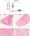
Results: The cell count in the decellularized menisci was 11 cells/mm² (SD = 13; 95% CI: 2-20), representing a significant reduction compared to the native meniscus samples, which had a cell count of 111 cells/mm² (SD = 42; 95% CI: 81-141; p < 0.01). Young's modulus of elasticity was 35.3 versus 36.8 MPa in the anterior region (p = 0.8), 32.6 versus 35.6 MPa in the central region (p = 0.7) and 36.5 versus 35.8 MPa in the posterior region (p = 0.9) for native versus decellularized samples, respectively.
Conclusions: This study demonstrated that the modified SDS-based decellularization protocol effectively decellularizes the human meniscus. Moreover, the decellularized tissue retained biomechanical properties comparable to those of native meniscus tissue. Tissue decellularization is a promising technique in regenerative medicine, enabling the use of scaffolds for tissue repair, particularly in applications such as meniscus transplantation following meniscectomy.
Level of evidence: Level III, controlled laboratory study.
Keywords: compressive biomechanical properties; decellularization; knee; meniscus tissue engineering; scaffolds.
© 2025 The Author(s). Journal of Experimental Orthopaedics published by John Wiley & Sons Ltd on behalf of European Society of Sports Traumatology, Knee Surgery and Arthroscopy.
Conflict of interest statement
The authors declare no conflicts of interest.
Figures


Similar articles
-
Prescription of Controlled Substances: Benefits and Risks.2025 Jul 6. In: StatPearls [Internet]. Treasure Island (FL): StatPearls Publishing; 2025 Jan–. 2025 Jul 6. In: StatPearls [Internet]. Treasure Island (FL): StatPearls Publishing; 2025 Jan–. PMID: 30726003 Free Books & Documents.
-
Can the Acetabular Labrum Be Reconstructed With a Meniscal Allograft? An In Vivo Pig Model.Clin Orthop Relat Res. 2024 Feb 1;482(2):386-398. doi: 10.1097/CORR.0000000000002860. Epub 2023 Sep 19. Clin Orthop Relat Res. 2024. PMID: 37732715 Free PMC article.
-
Tissue engineering and regenerative medicine strategies in meniscus lesions.Arthroscopy. 2011 Dec;27(12):1706-19. doi: 10.1016/j.arthro.2011.08.283. Epub 2011 Oct 21. Arthroscopy. 2011. PMID: 22019234
-
Meniscal Lesions in Multi-Ligament Knee Injuries.Indian J Orthop. 2024 Jul 11;58(9):1224-1231. doi: 10.1007/s43465-024-01217-0. eCollection 2024 Sep. Indian J Orthop. 2024. PMID: 39170649 Free PMC article.
-
In elite athletes with meniscal injuries, always repair the lateral, think about the medial! A systematic review.Knee Surg Sports Traumatol Arthrosc. 2023 Jun;31(6):2500-2510. doi: 10.1007/s00167-022-07208-8. Epub 2022 Nov 2. Knee Surg Sports Traumatol Arthrosc. 2023. PMID: 36319751 Free PMC article.
References
-
- Abdelgaied A, Stanley M, Galfe M, Berry H, Ingham E, Fisher J. Comparison of the biomechanical tensile and compressive properties of decellularised and natural porcine meniscus. J Biomech. 2015;48:1389–1396. - PubMed
-
- Baratz ME, Fu FH, Mengato R. Meniscal tears: the effect of meniscectomy and of repair on intraarticular contact areas and stress in the human knee: a preliminary report. Am J Sports Med. 1986;14(3):270–275. - PubMed
-
- Bellisari G, Samora W, Klingele K. Meniscus tears in children. Sports Med Arthrosc. 2011;19(1):50–55. - PubMed
LinkOut - more resources
Full Text Sources
Research Materials

