. 2025 Mar;8(3):218-228.
doi: 10.1038/s41929-025-01294-w. Epub 2025 Feb 6.

Iminium Catalysis in Natural Diels-Alderase

Affiliations

Iminium Catalysis in Natural Diels-Alderase

Zuodong Sun et al. Nat Catal. 2025 Mar.

Abstract

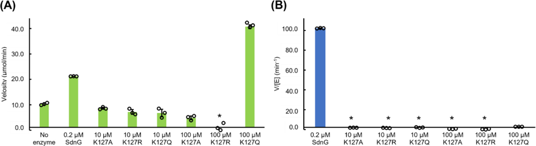
Iminium-catalyzed cycloaddition is one of the most prominent examples of organocatalysis, yet a biological counterpart has not been reported despite the wide-spread occurrence of iminium adducts in enzymes. Here, we present biochemical, structural, and computational evidence for iminium catalysis by the natural Diels-Alderase SdnG that catalyzes norbornene formation in sordarin biosynthesis. A Schiff base adduct between the ε-nitrogen of active site K127 and the aldehyde group of the enal dienophile was revealed by structural analysis and captured under catalytic conditions via borohydride reduction. This Schiff base adduct positions the substrate into near-attack conformation and decreases the transition state barrier of Diels-Alder cyclization by 8.3 kcal/mol via dienophile activation. A hydrogen bond network consisting of a catalytic triad is proposed to facilitate proton transfer required for iminium formation. This work establishes a new mode of catalysis for Diels-Alderases and points the way to the design of novel iminium based (bio)catalysts.


Figures

Extended Data Figure 1.
Examples of biocatalysis involving iminium enzyme-substrate adducts. (A) Naturally occurring Schiff base dependent enzymes. (B) Iminium catalysis by engineered enzymes.
Extended Data Figure 2.
The active sites of SdnG complexed with different ligands are nearly superimposable. (A) Overlay of active sites of SdnG-3NC (ivory) and SdnG-3C (gray). Noncovalently bound 3 and covalently bound 3 are shown in cyan and green respectively. Water molecules interacting with 3 and Y41 are omitted for clarity. (B) Overlay of active sites of SdnG-3NC (ivory) and SdnG-4NC (gray). 3 and 4 are shown in cyan and green respectively. (C) Overlay of active sites of SdnG-3C (ivory) and SdnG-7 (gray). Covalently bound 3 and adduct 7 are shown in cyan and green respectively.
Extended Data Figure 3.
K127 adducts corresponding to the rDA and Cope rearrangement products of intermediate 7 were observed in the crystal structure of SdnG with 4. (A) Active site of SdnG-7-5 (the rDA product of 7, chain C of SdnG-4, PDB 8YI8). Adducts 7 and 5 are shown in cyan and green respectively. Residues around 4 Å of the ligands are shown in ivory. The Polder omit map of 7–5 (contoured at 2.0 σ) is shown in blue mesh. Maps contoured at higher levels (2.5 σ and 3.0 σ) are shown in Supplementary Fig. 11. (B) Distance between diene and dienophile (highlighted in purple) of 5 in SdnG-7-5. Active site residues and 5 are shown in ivory and green respectively. Adduct 7 is omitted from the view for clarity. (C) Active site of SdnG-8 (the Cope rearrangement product of 7, PDB 8YHM). Adduct 8 is shown in cyan. The Polder omit map of 8 (blue mash) is contoured at 3.0 σ. Distances between atoms are shown in dashed lines. Bonds corresponding to the diene and the dienophile in 1 are highlighted in purple. Other coloring schemes are the same as in (A). (D) DFT calculated transition states of rDA and Cope rearrangement of 7. All energies are relative to the ground state energy of 5 (iminium form). Bonds corresponding to the diene and the dienophile in 1 are highlighted in blue in 5 and green in 8. The transition state structure of rDA (TS-5) is the same as that of the forward reaction (TS-4). But the barriers of the forward and reverse reactions differ significantly due to the large energy gap between 5 and 7.
Extended Data Figure 4.
Catalytic activity of SdnG and K127X mutants. All reactions were carried out for 1 min with 100 μM 1. Values and error bars are obtained from the average and standard deviation of three independent measurements (black circles) respectively (n=3). (A) Absolute rates of nonenzymatic and enzymatic DA cyclization of 1. Asterisks indicate no measurable substrate consumption during the course of the reaction. (B) Relative activity of SdnG and K127X variants normalized by enzyme concentration. All activities are shown relative to the rate acceleration of the DA reaction exhibited by the wild-type enzyme (WT, 100%). A 0% value (asterisks) indicates no rate acceleration compared to uncatalyzed DA reaction.
Extended Data Figure 5.
Effect of the concentration of 4 on formation of iminium 7 in SdnG and the H72A variant. SdnG or its mutational variants (5 μM) were mixed with varied amounts of 4 and the mixture was immediately treated with 20 mM NaBH4. The reaction was subsequently analyzed by UHPLC-HRMS and the deconvoluted ESI-MS spectra were shown in the figure. Increased concentration of 4 promotes imine formation of SdnG but not the H72A variant. The result suggests that ligand binding is rate-determining for iminium formation in SdnG but not in the H72A variant. Therefore, diminished iminium formation in H72A is not a result of compromised ligand binding.
Figure 1.
Figure 1.
SdnG is proposed to be a Diels-Alderase that performs iminium catalysis. (A) Macmillan’s example of iminium-catalyzed cycloaddition for norbornene synthesis. (B) Proposed iminium-catalyzed DA reaction by SdnG. Compound 1 is the substrate of SdnG, while 4 is the product. An active site lysine (in blue) is proposed to be the catalytic residue that forms the iminium adduct. Compound 3 is a substrate analog derived from 6 and is used in X-ray crystallography. 6 is recovered as a shunt product from A. nidulans heterologous expression and is reduced from 1 by cellular reductases, likely using NADPH as a reductant. GGPP: geranylgeranyl pyrophosphate. (C) DFT calculated intramolecular DA transition states of 1 and its adducts with a lysine (modeled as methyl amine). TS-1, DA transition state (TS) of compound 1 (aldehyde); TS-2, DA TS of imine adduct of 1; TS-3, DA TS of imine adduct of 1 hydrogen bonded to a serine (modeled as methanol); TS-4, DA TS of the iminium adduct of 1. All energies (ΔG) are relative to the ground state energy of 1 or its derivatives. The distances between atoms are shown in Å.
Figure 2
Figure 2
Crystal structures of SdnG with substrate analog 3 and product 4. (A) Backbone structure of SdnG-3NC (PDB 8YHG). The two monomers of SdnG are colored in ivory and pink respectively. The active site cavity is represented by gray surface and 3 is shown in cyan sticks. (B) Active site structure of SdnG-3NC. Compound 3 and residues around 4 Å are shown in cyan and ivory respectively. The Polder omit map of 3 (contoured at 3.0 σ) is shown in blue mesh. Water molecules are shown in red spheres. Bonds corresponding to the diene and the dienophile in 1 are highlighted in purple. Distances between atoms are shown in dashed lines. The same coloring scheme is used throughout Figure 2. (C) Active site structure of SdnG-3C (PDB 8YJ4). The Polder omit map of the K127-3 adduct is contoured at 3.0 σ. Water molecules interacting with 3 and Y41 are omitted for clarity. (D) Active site of SdnG-4NC (chain A of SdnG-4, PDB 8YI8). The Polder omit map of 4 is contoured at 3.0 σ. (E) Active site of SdnG-7 (chain B of SdnG-4, PDB 8YI8). The Polder omit map of adduct 7 is contoured at 3.0 σ. Water molecules interacting with 7 and Y41 are omitted for clarity. (F) Active site hydrogen bond network in SdnG-7.
Figure 3
Figure 3
Biochemical evidence of iminium catalysis in SdnG. (A) Conservation of featured residues in SdnG homologs shown in sequence logos. (B) Formation of Schiff base adducts between SdnG or its K127A variant and compounds 3 or 4 under catalytic conditions. Enzymes were mixed with the indicated ligand and the mixture was immediately treated with 20 mM NaBH4. The reaction was subsequently analyzed by UHPLC-HRMS and the deconvoluted ESI-MS spectra were shown for the following reactions: i) 5 μM SdnG without ligand; ii) 5 μM SdnG mixed with 5 μM 3; iii) 5 μM SdnG mixed with 5 μM 4; iv) 5 μM K127A without ligand; v) 5 μM K127A mixed with 50 μM 4. (C) In vitro activity of SdnG and its mutational variants. All activities are shown relative to the rate acceleration of the DA reaction exhibited by the wild-type enzyme (WT, 100%). A 0% value (asterisks) indicates no rate acceleration compared to uncatalyzed DA reaction. Values and error bars are obtained from the average and standard deviation of three independent measurements (black circles) respectively (n=3). (D) Effect of mutations of the active site hydrogen bond network on the formation of Schiff base adduct. SdnG or its mutational variants (5 μM) were mixed with compound 4 (5 μM) and the mixture was immediately treated with 20 mM NaBH4. The reaction was subsequently analyzed by UHPLC-HRMS and the deconvoluted ESI-MS spectra were shown in the figure.
Figure 4.
Figure 4.
Proposed mechanism of SdnG. Enzyme (E), substrate (S) and product (P) represent SdnG, 1, and 4 respectively. Important polar and nonpolar interactions are shown in dashed lines. Interactions involving Y41 are omitted for clarity. Nonproductive conformation of 1 is shown in orange in the ES complex.

References

    1. MacMillan DWC The advent and development of organocatalysis. Nature 455, 304–308 (2008). - PubMed
    1. Ahrendt KA, Borths CJ & MacMillan DWC New Strategies for Organic Catalysis: The First Highly Enantioselective Organocatalytic Diels−Alder Reaction. J. Am. Chem. Soc 122, 4243–4244 (2000).
    1. Gefflaut T, Blonski C, Perie J. & Willson M. Class I aldolases: Substrate specificity, mechanism, inhibitors and structural aspects. Prog. Biophys. Mol. Biol 63, 301–340 (1995). - PubMed
    1. Fan P-H, Sato S, Yeh Y-C & Liu H. Biosynthetic Origin of the Octose Core and Its Mechanism of Assembly during Apramycin Biosynthesis. J. Am. Chem. Soc 145, 21361–21369 (2023). - PMC - PubMed
    1. Piersen CE, McCullough AK & Lloyd RS AP lyases and dRPases: commonality of mechanism. Mutat. Res. Repair 459, 43–53 (2000). - PubMed

LinkOut - more resources