. 2024 Dec:109:451-486.
doi: 10.3114/sim.2024.109.07. Epub 2025 Oct 4.

Examination of mycoparasites reveals a new type of host-parasite interface and rearranges the taxonomy of Occultifur and Microsporomyces (Cystobasidiomycetes, Basidiomycota)

Affiliations

Examination of mycoparasites reveals a new type of host-parasite interface and rearranges the taxonomy of Occultifur and Microsporomyces (Cystobasidiomycetes, Basidiomycota)

N Schoutteten et al. Stud Mycol. 2024 Dec.

Abstract

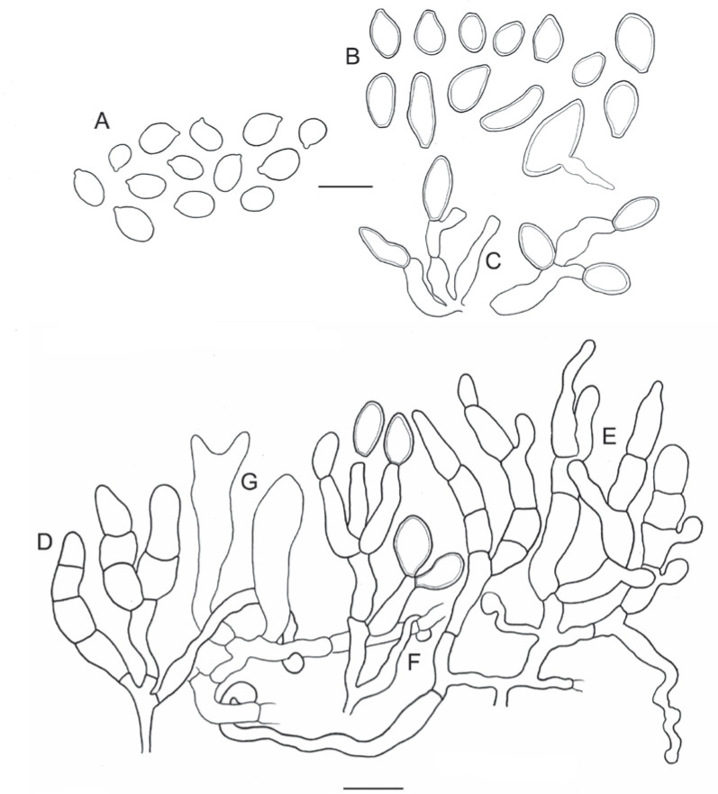
The present study investigates the species boundaries, evolutionary relationships, and host-parasite interfaces of dimorphic mycoparasites that were previously assigned to Achroomyces soranus, Occultifur internus, and Platygloea mycophila based on morphological similarities. Our comparison of recently collected and cultivated samples with the type specimens of A. soranus and P. mycophila shows that both groups are species complexes, of which the taxa can be differentiated based on morphological and ecological characters. By integrating the results of a seven-locus dataset (SSU, LSU, ITS, RPB1, RPB2, TEF1, and mitochondrial CYT-B) and detailed micromorphological comparisons of the investigated specimens, we show for the first time that these three groups of mycoparasites belong to Cystobasidiomycetes (Pucciniomycotina). We applied a polyphasic species concept involving morphology, phylogeny, and ecology to delineate and circumscribe these and new genera. The genus Occultifur comprises six species. Occultifur internus and the newly proposed O. cerinomycicola are intrahymenial mycoparasites producing haustorial cells and establishing fusion pore interaction with their Dacrymycetous host. Based on microscopical examination, we show that Achroomyces soranus is a member of the genus Occultifur. Based on the molecular phylogenetic reconstruction, we found that three lichen-associated fungi which are only known from a yeast morph are nested within Occultifur, i.e. Lichenozyma pisutiana, Microsporomyces cladoniae, and M. wangii. The genus Obvidator is newly introduced for three mycoparasitic species inhabiting members of the corticioid genus Peniophora (Russulales, Agaricomycetes) and causing gall-like malformations of the host basidiome. Microscopic investigation shows that Platygloea mycophila is a member of this genus. Obvidator species display a yet undiscovered type of host-parasite interface, in which the mycoparasites produce short protrusions on their hyphae adjacent to the host hyphae. The lysis of the host cell wall takes place at points of contact with parasite protrusions, but no rupture of the host plasma membrane or nanometer-fusion pore formation was observed. The updated Cystobasidiomycetes phylogeny obtained in this study by including mycoparasites showed that the genera Occultifur and Microsporomyces as currently circumscribed are polyphyletic. To resolve this polyphyly, we introduce two new genera, i.e. Cystastrum and Millanizyma, and recombine species comprising the Occultifur externus clade and a clade consisting of Microsporomyces bloemfonteinensis and M. cladoniophilus, respectively. Taxonomic novelties: New genera: Cystastrum Schoutteten & Yurkov, Millanizyma Schoutteten & Yurkov, Obvidator Schoutteten. New species: Obvidator incarnatae Schoutteten & Yurkov, Obvidator quercinae Schoutteten & Yurkov, Occultifur cerinomycicola Schoutteten, Enzlin & Yurkov. New combinations: Cystastrum brasiliense (F.C.O. Gomes et al.) Schoutteten, Cystastrum externum (J.P. Samp. et al.) Schoutteten & Yurkov, Cystastrum kilbournense (Kurtzman & Robnett ex Denchev & T. Denchev) Schoutteten & Yurkov, Cystastrum mephitis (Zalar et al.) Schoutteten & Yurkov, Cystastrum tropicale (Khunnamw. et al. ex Denchev & T. Denchev) Schoutteten & Yurkov, Cystastrum plantarum (Khunnamw. et al.) Schoutteten & Yurkov, Millanizyma bloemfonteinensis (C.H. Pohl et al.) Schoutteten & Yurkov, Millanizyma cladoniophila (N.H. Nguyen et al.) Schoutteten & Yurkov, Occultifur cladoniae (Kachalkin et al.) Schoutteten, Occultifur pisutianus (Černajová & Škaloud) Schoutteten, Obvidator mycophilus (Burds. & Gilb.) Schoutteten, Occultifur soranus (Hauerslev) Schoutteten, Occultifur wangii (Kachalkin et al.) Schoutteten. Citation: Schoutteten N, Yurkov A, Spirin V, Savchenko A, Aime MC, Begerow D, Verbeken A (2024). Examination of mycoparasites reveals a new type of host-parasite interface and rearranges the taxonomy of Occultifur and Microsporomyces (Cystobasidiomycetes, Basidiomycota). Studies in Mycology 109: 451-486. doi: 10.3114/sim.2024.109.07.

Keywords: Basidiomycota; dimorphism; fungal systematics; haustorial cells; host-parasite interaction; molecular phylogeny; mycoparasitism; new taxa; taxonomy; transmission electron microscopy; yeast.


Conflict of interest statement

The authors declare that there is no conflict of interest.

Figures

Fig. 1
Fig. 1
Phylogenetic relationships of mycoparasites in Cystobasidiomycetes based on a seven-locus ML tree reconstruction. Species names in bold indicate taxonomic novelties. By default, included species are only known from a yeast morph. F indicates that both the yeast and filamentous morphs are known. M refers to mycoparasitic species. Clades comprising mycoparasites which are investigated in this study are indicated with yellow boxes. Vertical lines represent the highest described taxon available for species in the tree (family or order). Numbers of branches indicate ultrafast bootstrap values. Naohideales is used as outgroup.
Fig. 1
Fig. 1
Phylogenetic relationships of mycoparasites in Cystobasidiomycetes based on a seven-locus ML tree reconstruction. Species names in bold indicate taxonomic novelties. By default, included species are only known from a yeast morph. F indicates that both the yeast and filamentous morphs are known. M refers to mycoparasitic species. Clades comprising mycoparasites which are investigated in this study are indicated with yellow boxes. Vertical lines represent the highest described taxon available for species in the tree (family or order). Numbers of branches indicate ultrafast bootstrap values. Naohideales is used as outgroup.
Fig. 2
Fig. 2
Occultifur soranus comb. nov. (C-F-39334) line drawings. A. Basidiospores. B. Thick-walled conidia, note one germinating conidium. C. Conidiogenous cells giving rise to thick-walled conidia. D. Cluster of transversally septate basidioles. E. Cluster of transversally 3-septate basidia. F. Haustorial cell with globular apex curving around host hypha. G. Host basidiole and basidium. Scale bars = 10 μm.
Fig. 3
Fig. 3
Occultifur soranus comb. nov. (C-F-39334). A. Host basidiomata, arrowheads indicate host basidiomata that were investigated and found to contain the intrahymenial parasite. B. Conidiogenous cells and conidia. Double arrowhead indicates a conidiogenous cell directly developing a conidium. C. Thick-walled conidium and a cluster of basidioles showing a variable number of transverse septa. D. Cluster of two 3-septate basidia, with variable number of outgrowing sterigmata. E. Detached 3-septate basidium with a sterigma growing from each basidial compartment. F. Double arrow indicates a conidiogenous cell bearing a conidium. G, H. Host-parasite interface, Pa = parasite, Ho = host, arrowheads show globose apices of the haustorial cells attaching to host hyphae. Scale bars: A = 1 cm; B–H = 10 μm.
Fig. 4
Fig. 4
Occultifur cerinomycicola sp. nov. (ENZ 22-005) line drawings. A. Thick-walled conidia. B. Basidiospores. C. Thick-walled conidiogenous cells giving rise to conidia. D. Cluster of transversally-septate basidioles. E. Transversally 3-septate basidium. F. Haustorial cell attaching to a host hypha. G. Host basidia. Scale bars = 10 μm.
Fig. 5
Fig. 5
Occultifur cerinomycicola sp. nov. (ENZ 22-005). A. Host basidiomata. B. Cluster of transversally-septate basidioles and basidia, developing from a thick-walled hypha. C. Transversally 3-septate basidim with two outgrowing sterigmata, arrowhead shows a curving haustorial cell around the basidium base. D. Collapsing basidium with outgrowing sterigmata, arrowheads indicate two haustorial cells. E. Cluster of conidiogenous cells (double arrowheads) giving rise to conidia. F. Host-parasite interface, Pa = parasite, Ho = host, arrowheads show haustorial cells. Scale bars: A = 1 cm; B–F = 10 μm.
Fig. 6
Fig. 6
Obvidator incarnatae sp. nov. (MJD 20-030) line drawings. A. Basidiospores, note some spores germinating by hypae. B. Transversally 2-septate basidium with thick-walled, swollen probasidium and two outgrowing sterigmata. C. Cluster of terminal cells. D. Thick-walled parasite hypha producing interaction protrusions. E. Gloeocystidium of the host adjacent to host protrusions. Scale bars = 10 μm.
Fig. 7
Fig. 7
Obvidator incarnatae sp. nov. (MJD 20-030). A. Host basidiome showing infection symptoms visible as concolorous to salmon-coloured galls. Photo courtesy of M. Jagers. B. Cluster of transversally 2-septate basidia, p = probasidium, s = sterigma, 1 = first basidial compartment, 2 = second basidial compartment. C. Thick-walled hyphae giving rise to terminal cells with variable shapes. D. Basidiospores. E, F. Host-parasite interface, arrowheads show protrusions of mycoparasite cells that attach or point to adjacent host cells. Scale bars: A = 1 cm; B–F = 10 μm.
Fig. 8
Fig. 8
Obvidator mycophilus comb. nov. (RLG-10056) line drawings. A. Basidiospores. B. Transversally 2-septate basidium with thick-walled, swollen probasidium and two outgrowing sterigmata. C. Cluster of terminal cells with variable morphology. D. Thick-walled parasite hypha producing interaction protrusions. E. Clamped host cell adjacent to host protrusions. Scale bars = 10 μm.
Fig. 9
Fig. 9
Obvidator mycophilus comb. nov. (RLG-10056). A. Host basidiome showing infection symptoms visible as yellowish galls (arrowheads) B. Transversally 2-septate basidium with two outgrowing sterigmata, the probasidium is swollen. C. Thick-walled hyphae giving rise to terminal cells with variable shapes. D. Basidiospores, one spore germinates by an outgrowing hypha. E–G. Host-parasite interface, Pa = parasite, Ho = host, arrowheads show protrusions of mycoparasite cells that attach or point to adjacent host cells. Scale bars = 10 μm.
Fig. 10
Fig. 10
Obvidator quercinae sp. nov. (NS 23-005) line drawings. A. Basidiospores, note one repetitive spore engaging in secondary spore production and multiple spores germinating by hyphae. B. Cluster of transversally 2-septate basidia with thick-walled, slightly swollen probasidia, and outgrowing sterigmata. C. Cluster of terminal cells with variable morphology, note one bifurcating terminal cell. D. Thick-walled parasite hypha producing interaction protrusions. E. Clamped host cell adjacent to host protrusions. Scale bars = 10 μm.
Fig. 11
Fig. 11
Obvidator quercinae sp. nov. (from specimens NS 19-450 (A) and NS 23-005 (B–G)). A. Host basidiome showing infection symptoms visible as concolorous galls. Photo courtesy of J. Heilmann-Clausen. B. Cluster of terminal cells, basidium and basidiospores. C, G. Transversally two-septate basidium, p = probasidium, s = sterigma, 1 = first basidial compartment, 2 = second basidial compartment. D. Basidiospores. E, F. Host-parasite interface, Pa = parasite, Ho = host, arrowheads show protrusions of mycoparasite cells that attach or point to adjacent host cells. Scale bars: A = 1 cm; B–G = 10 μm.
Fig. 12
Fig. 12
Transmission electron microscopy of the host-parasite interface of Obvidator quercinae sp. nov. and its host Peniophora quercina. P = mycoparasite, H = host. A. Longitudinally sectioned parasite protrusion making contact with a host cell. B. Magnification of A, arrowheads indicate the rupturing cell wall of the host. C. Transversally sectioned parasite protrusion making contact with a host cell. D. Magnification of C, arrowheads indicate the rupturing cell wall of the host, double arrowhead indicates the intact plasmamembrane of the host cell. E. Irregularly shaped parasite protrusion making contact with a host cell. F. Magnification of E, arrowheads indicate the rupturing cell wall of the host, double arrowhead indicates the intact plasmamembrane of the host cell.
Fig. 13
Fig. 13
Transmission electron microscopy of the host-parasite interface of Obvidator quercinae sp. nov. and its host Peniophora quercina. P = mycoparasite, H = host. A. Host cell surrounded by parasite protrusions. B. Magnification of A, arrowheads indicate the rupturing cell wall of the host, double arrowhead indicates the intact plasmamembrane of the host cell. C. Irregularly shaped parasite protrusion neighbouring two host cells, arrowhead indicates rupturing of the cell wall of the left host cell. D. Magnification of C, between the double arrowheads is a structure that resembles a nanopore fusion channel. Note that in C and D the cytoplasm of the host cells appears degraded. E. Parasite protrusion adjacent to a host cell with cytoplasm that appears degraded, arrowheads indicate the rupturing host cell wall. F. Pyriform parasite protrusion making contact with a host cell, double arrowhead indicates the intact plasmamembrane of the host, note the electron transparent layer surrounding the protrusion.
Fig. 14
Fig. 14
Culture characteristics of the isolated mycoparasites. A. Yeast colonies of Occultifur cerinomycicola sp. nov. (DSM 115928). B. Yeast cells of O. cerinomycicola (DSM 115928). C. Yeast colonies of Obvidator incarnatae sp. nov. (DSM 116223) D. Yeast cells of Ob. incarnatae (DSM 116223). E. Yeast colonies of Obvidator quercinae sp. nov. (DSM 117925). F. Yeast cells of Ob. quercinae (DSM 117925). Yeast cells in B, D, and F are prepared in 10 % glycerin and are visualised with DIC microscopy. Scale bars: A, C, E = 2 mm; B, D, F = 10 μm.

References

    1. Aime MC, Toome M, McLaughlin DJ. (2014). Pucciniomycotina. In: The Mycota VII. Systematics and evolution. Part A. (McLaughlin DJ, Spatafora JW, eds). Springer, Germany: 271–294.
    1. Akulov OY, Fomenko MI, Khudych AS. et al. (2022). The first find of Naohidea sebacea (Naohideales, Basidiomycota) in Ukraine. Ukrainian Botanical Journal 79(5): 308–313.
    1. Anslan S, Mikryukov V, Armolaitis K. et al. (2021). Highly comparable metabarcoding results from MGI-Tech and Illumina sequencing platforms. PeerJ 9: e12254. - PMC - PubMed
    1. Bai FY, Cai Y, Wang QM. et al. (2004). Rhodotorula oryzae sp. nov., a novel basidiomycetous yeast species isolated from paddy rice. Antonie van Leeuwenhoek 86: 295–299. - PubMed
    1. Bai F, Liu Y, Li N. et al. (2016). Microsporomyces hainanensis sp. nov., isolated from hybrid rice (Oryza sativa L.) seeds. Current Microbiology 73: 569–573. - PubMed

LinkOut - more resources