. 2025 Nov:61:102512.
doi: 10.1016/j.tranon.2025.102512. Epub 2025 Aug 28.

An anti-CEA affibody showing high-definition staining in human pancreatic cancer tissue sections and selective tumor targeting in vivo

Affiliations

An anti-CEA affibody showing high-definition staining in human pancreatic cancer tissue sections and selective tumor targeting in vivo

Johan Nilvebrant et al. Transl Oncol. 2025 Nov.

Abstract

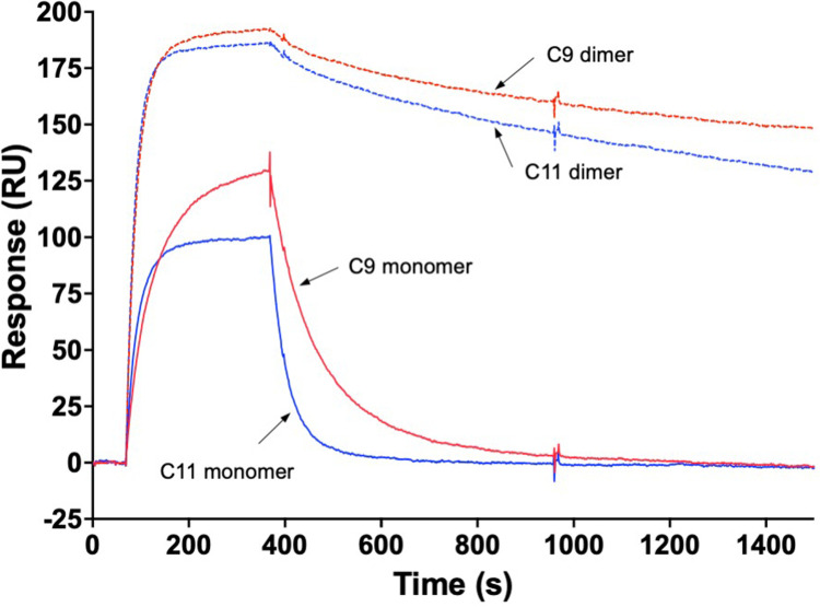
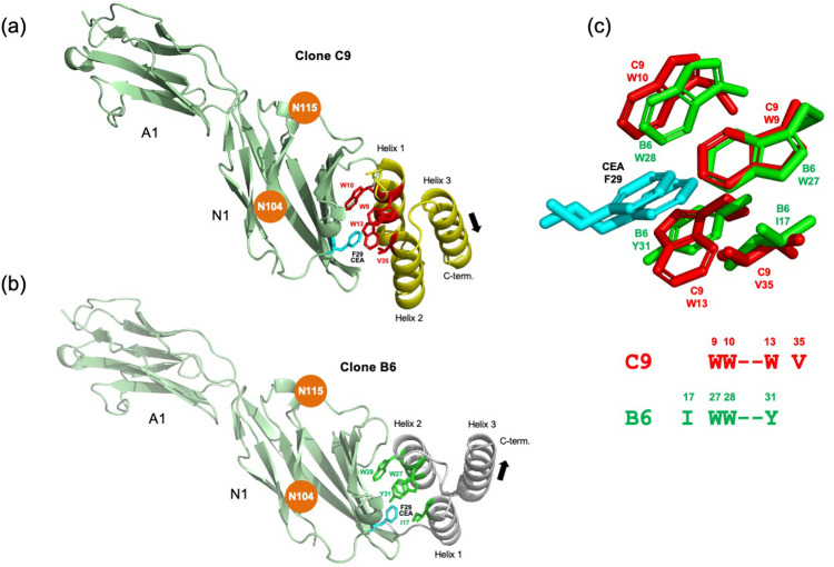
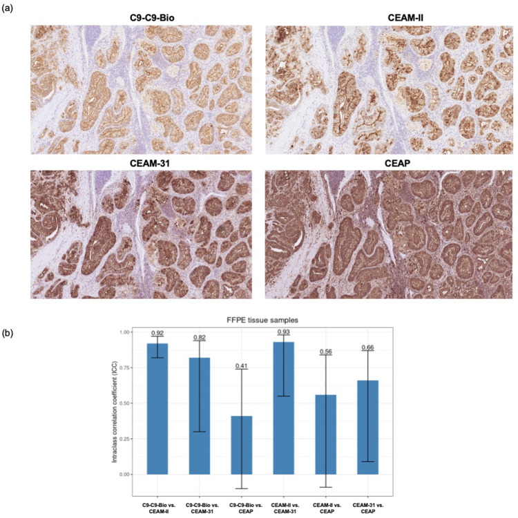
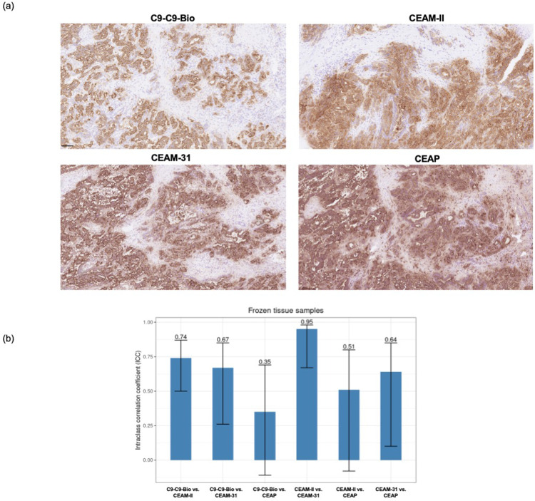
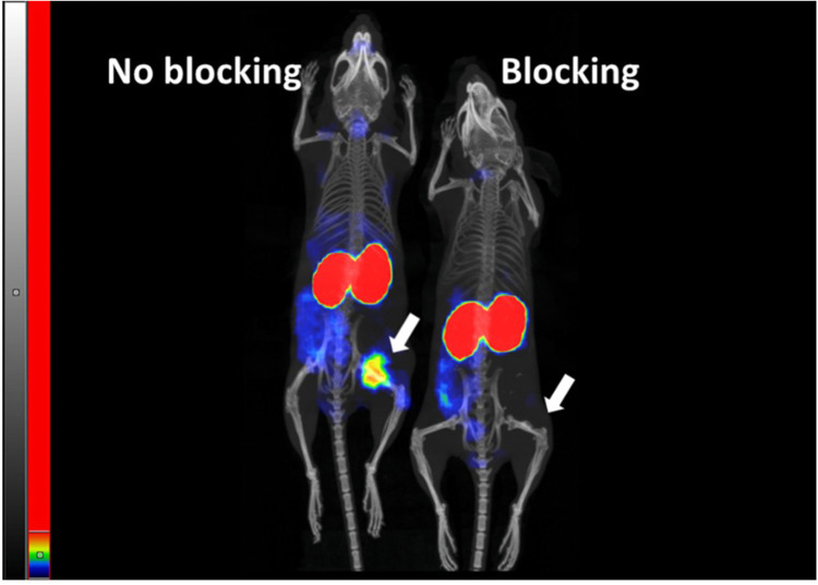
We report development and characterization of small non-immunoglobulin affibody affinity proteins directed to the highly glycosylated human carcinoembryonic antigen-related adhesion molecule 5 (CEACAM5, CEA), and their use in immunohistochemical (IHC) analyses of human pancreatic cancer samples and for in vivo tumor imaging. A total of nineteen unique anti-CEA affibodies were identified from large phage display libraries constructed using combinatorial protein engineering of a small 58 amino acid three-helix bundle protein domain. Molecular modeling suggested that all enriched clones share a binding surface with several clustered tryptophan residues interacting with a hydrophobic patch in the N1 domain of CEA centered around a phenylalanine residue. One variant, designated as C9, exhibited the highest affinity in biosensor analyses and was reformatted into a 15 kDa homodimer expressed in Escherichia coli. The biotinylated form, C9-C9-Bio, was evaluated for its IHC performance on matched frozen and formalin-fixed, paraffin-embedded (FFPE) sections of human pancreatic cancer samples (n = 7). Compared to clinical-grade monoclonal antibodies II-7 and CEA31, as well as a polyclonal reagent, C9-C9-Bio demonstrated highly sensitive CEA detection with minimal background staining. Statistical analyses including intraclass correlation and Bland-Altman assessments revealed excellent agreement between C9-C9-Bio and the two monoclonal antibodies in FFPE tissue samples. Further, a 99mTc[Tc]-labeled C9-C9 construct showed CEA-dependent binding to human cancer cell lines in vitro, and selectively bound to CEA-expressing BxPC3 xenografts in mice when investigated as a tracer for in vivo imaging, allowing for a visualization of tumors after four hours. In summary, these findings highlight the potential use of the easily produced CEA-binding C9 affibody for various clinical applications, including IHC and medical imaging, and as a targeting moiety for directing various therapeutic modalities to CEA-expressing tumors.

Keywords: Adenocarcinoma; Affibody; Affinity; CEA; CEA31; CEACAM5; Carcinoembryonic antigen; Clinical diagnostics; HRP; IHC; II-7; Immunohistochemistry; In vivo imaging; Pancreatic cancer; Phage display; Xenograft.


Conflict of interest statement

Declaration of competing interest The authors declare the following financial interests/personal relationships which may be considered as potential competing interests: The new anti-CEA affinity proteins described in the manuscript are subject of a patent application, involving the following inventors: JN, CFM, HS, AA, MB and PÅN.

Figures

Fig. 1
Fig. 1
Schematic description of the main proteins included in this study. (a) Overall three-dimensional structure of the 58 aa affibody scaffold, showing the three-helix bundle organization. The 14 positions in helices 1 and 2 that are subjected to combinatorial randomization to construct libraries are highlighted in red and numbered. (b) Molecular model of the seven-domain CEA 3D-structure 1E07.pdb previously validated using solution scattering data [7]. (c) Schemes of the different affibody formats used within the present study including (i) His6-affibody-ABD, where ABD is a 46 aa serum albumin binding domain, (ii) affibody-affibody-His6, (iii) His6-affibody-affibody-Cys, (iv) a dimeric anti-CEA affibody C9 construct conjugated to maleimide-PEG-biotin (C9-C9-Bio) and (v) a dimeric anti-CEA affibody C9 construct containing an EYEC extension allowing for site-specific labeling with [99mTc]Tc.
Fig. 2
Fig. 2
Homodimeric affibody constructs bind with significantly higher apparent affinity to CEA compared to monomers. Proteins were injected at 200 nM concentrations over a sensor chip surface containing immobilized human CEA protein. (a) Binding of monomeric C9-His6 and C11-His6 proteins, and (b) binding of homodimeric His6-C9-C9-ABD and His6-C11-C11-ABD proteins to CEA. Note the significantly slower off-rate kinetics for homodimeric affibody constructs, via avidity effects.
Fig. 3
Fig. 3
Biosensor binding competition experiments reveal the high specificity of dimeric affibody constructs to CEA. Binding of either free CEA or CEA mixed with a 10-fold molar excess of either C9-C9-His6, C9-C9-His6 or anti-CEA MFE-23 scFv proteins to a sensor chip surface containing immobilized C9-C9-His6, demonstrated that all three analytes blocked binding of CEA to C9-C9-His6. Inset: Analysis of the binding of a concentration series (100–1 nM, 3-fold dilutions) of free anti-CEA MFE-23 scFv to a sensor chip surface containing immobilized CEA.
Fig. 4
Fig. 4
Substitution of specific tryptophan residues in C9 abrogates binding to CEA. The binding capacity of 14 alanine substituted C9 variants was compared to parental C9 (C9 wild type (WT), bold red trace). The binding of each monovalent affibody-His6 protein to immobilized CEA is presented. The same concentration of 200 nM was used for all variants. Variants N18A, S14A, D25A, Q27A, V28A and K24A showed essentially retained binding affinity, whereas variants H32A, V35A, V17A, K31A and F11A displayed markedly reduced binding responses. The absence of detectable binding at 200 nM for the three variants W9A, W10A and W13A was confirmed by a second analysis using 2000 nM (inset).
Fig. 5
Fig. 5
AlphaFold3 models of affibody-CEA complexes. AlphaFold3-advanced [1,27] was used to model complexes between CEA and either of two anti-CEA affibodies. (a)Affibody C9:CEA complex. Showing one of several generated models suggesting that a region at the very end of domain N1 contained the epitope for the C9 affibody. Centrally positioned in the binding interface is a phenylalanine 29 (F29) from CEA, whose side chain appears to interact with all three aromatic side chains of the W9-W10-W13 cluster in helix 1, and V35 in helix 2 of the C9 affibody. The proposed epitope is also free from postulated N-glycosylations, of which the closest are predicted to reside on positions N104 and N115 (highlighted in brown) at a certain distance from the epitope. (b)Affibody B6:CEA complex. The clone B6 was also analysed in the same manner. This clone also contains an aromatic cluster, W27-W28-Y32, but located in helix 2 which, together with I17 located in helix 1 also appears to be involved in binding to the side chain of F29 in CEA. Notably, the affibody is orientated in the opposite direction to align the cluster of clone B6 located on helix 2 which is anti-parallel to helix 1. (c) Alignment of aromatic clusters of C9 and B6. An alignment of the models of the aromatic clusters in C9 and B6, together with the F29 of CEA, shows a near perfect pairwise overlap of sidechains involved. Also shown is the alignment of the aliphatic residues V35 (C9) and I17 (B6), both appearing to contribute to the hydrophobic binding interface.
Fig. 6
Fig. 6
The homodimeric C9-C9 affibody allows for unambiguous detection of CEA in FFPE samples. (a) Representative photomicrograph of CEA detection with the dimeric C9-C9-Bio reagent and the clinical-grade monoclonal and polyclonal antibodies. (b) Intraclass correlation coefficients (ICC) as measurement of agreement in CEA detection scoring between the dimeric C9-C9-Bio reagent and the monoclonal and polyclonal antibodies.
Fig. 7
Fig. 7
CEA detection in frozen samples using the homodimeric C9-C9 affibody. (a) Representative photomicrograph of CEA detection with the dimeric C9-C9-Bio reagent and the clinical-grade monoclonal and polyclonal antibodies. (b) Intraclass correlation coefficients (ICC) as measurement of agreement in CEA detection scoring between the dimeric C9-C9-Bio reagent and the monoclonal and polyclonal antibodies.
Fig. 8
Fig. 8
Visualization of CEA-dependent uptake of [99mTc]Tc-C9-C9 in human cancer xenografts in mice (color). The animals were injected with the same activity of [99mTc]Tc-C9-C9 and the image of both animals was acquired 4 h after injection. Maximum Intensity Projection (MIP) is presented. The threshold of the linear color intensity scale is adjusted to the first red pixel in BxPC3 xenograft with high CEA expression.
Fig. 9
Fig. 9
Visualization of saturable uptake of [99mTc]Tc-C9-C9 in human cancer BxPC3 xenografts in mice. The animals were injected with the same activity of [99mTc]Tc-C9-C9 and the image of both animals was acquired 4 h after injection. Pre-injection of a large excess of non-labelled C9-C9-EYEC was used in the blocking experiment. Maximum Intensity Projection (MIP) is presented. The threshold of the linear color intensity scale is adjusted to the first red pixel in BxPC3 xenograft in the animal with non-blocked tumor.

References

    1. Abramson J., Adler J., Dunger J., Evans R., Green T., Pritzel A., Ronneberger O., Willmore L., Ballard A.J., Bambrick J., Bodenstein S.W., Evans D.A., Hung C.C., O'Neill M., Reiman D., Tunyasuvunakool K., Wu Z., Zemgulyte A., Arvaniti E., Beattie C., Bertolli O., Bridgland A., Cherepanov A., Congreve M., Cowen-Rivers A.I., Cowie A., Figurnov M., Fuchs F.B., Gladman H., Jain R., Khan Y.A., Low C.M.R., Perlin K., Potapenko A., Savy P., Singh S., Stecula A., Thillaisundaram A., Tong C., Yakneen S., Zhong E.D., Zielinski M., Zidek A., Bapst V., Kohli P., Jaderberg M., Hassabis D., Jumper J.M. Accurate structure prediction of biomolecular interactions with AlphaFold 3. Nature. 2024 - PMC - PubMed
    1. Ahlgren S., Andersson K., Tolmachev V. Kit formulation for 99mTc-labeling of recombinant anti-HER2 affibody molecules with a C-terminally engineered cysteine. Nucl. Med. Biol. 2010;37:539–546. - PubMed
    1. Altena R., Buren S.A., Blomgren A., Karlsson E., Tzortzakakis A., Brun N., Moein M.M., Jussing E., Frejd F.Y., Bergh J., Tran T.A., Hartman J., Axelsson R. Human epidermal growth factor receptor 2 (HER2) PET imaging of HER2-low breast cancer with [(68)Ga]Ga-ABY-025: results from a pilot study. J. Nucl. Med. 2024;65:700–707. - PubMed
    1. Baker M. Reproducibility crisis: blame it on the antibodies. Nature. 2015;521:274–276. - PubMed
    1. Ballehaninna U.K., Chamberlain R.S. Biomarkers for pancreatic cancer: promising new markers and options beyond CA 19-9. Tumour. Biol. 2013;34:3279–3292. - PubMed