. 2025 Aug 29;16(1):8074.
doi: 10.1038/s41467-025-63462-7.

NEXN protects against vascular calcification by promoting SERCA2 SUMOylation and stabilization

Affiliations

NEXN protects against vascular calcification by promoting SERCA2 SUMOylation and stabilization

Wenjie Guo et al. Nat Commun. .

Abstract

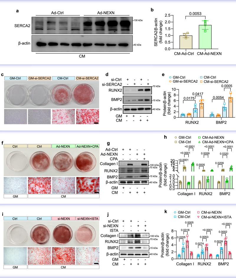
Vascular calcification, a key risk factor for cardiovascular diseases, is driven by the phenotypic transition of vascular smooth muscle cells from a contractile to an osteogenic phenotype. NEXN, a protein highly associated with heart function, has also been implicated as a potential susceptibility factor in the development of coronary artery disease, but its role in the progression of vascular calcification remains unclear. In this study, multi-transcriptomics analysis and various animal models of male mice were used to explore the cell-specific roles and molecular mechanisms of NEXN in vascular calcification. Here, we show that vascular smooth muscle cell-specific NEXN knockout exacerbates calcification, while NEXN overexpression alleviates it. Mechanistically, NEXN interacts with SERCA2, enhancing its SUMOylation, stability, and function, thereby protecting against calcification. These findings suggest potential therapeutic strategies by targeting NEXN-SERCA2 interactions or enhancing SERCA2 SUMOylation to prevent vascular calcification and its complications.


Conflict of interest statement

Competing interests: The authors declare no competing interests.

Figures

Fig. 1
Fig. 1. Identification of NEXN as a high-confidence gene associated with phenotypic transformation of VSMCs and vascular calcification.
a Volcano plot of DEGs (statistics in Limma). b Clustering of single-cell transcriptomes (GSE131780). c The expression of RUNX2, SPP1, SOX9 and ACAN in VSMCs. d Venn diagram showing intersecting genes. e The expression of NEXN in VSMCs. f MR analysis of association between artery NEXN mRNA expression and predisposition of coronary artery calcification (wald ratio method with statistical significance assessed via Wald ratio tests implemented in TwoSampleMR). g Representative western blot analysis of NEXN protein expression in heart, liver, kidney, aorta, skeletal muscle, lung, and brain tissue from male and female C57BL/6 mice (n = 4 biological replicates). h Representative immunofluorescence images showing NEXN (green) and RUNX2 (red) in control human aorta (No calcification) and calcified human aorta (With calcification) (n = 3 biological replicates). Scale bar: 50 µm.
Fig. 2
Fig. 2. NEXN expression was downregulated during vascular calcification in vitro and in vivo.
a Alizarin red staining of human VSMCs cultured for 7 days in GM or CM. Scale bar: 500 μm. b Quantitative analysis of alizarin red by microplate reader obtained from (a) (n = 4 biological replicates; error bar: mean (centre) ± standard deviation; statistical significance was assessed using two-sided and unpaired t-test). c Quantitative analysis of calcium content in human VSMCs cultured for 7 days in GM or CM (n = 4 biological replicates; error bar: mean (centre) ± standard deviation; statistical significance was assessed using two-sided and unpaired t-test). d Representative immunofluorescence for NEXN (green) and RUNX2 (red) in human VSMCs cultured for 7 days in GM or CM. Scale bar: 25 μm. e, f Western blot and quantification of the protein expression of NEXN, osteogenic markers Collagen I, RUNX2 and BMP2 in human VSMCs cultured for 7 days in GM or CM (n = 4 biological replicates; error bar: mean (centre) ± standard deviation; statistical significance was assessed using two-sided and unpaired t-test). g Alizarin red staining of the entire aortas of male C57BL/6 mice subjected to VitD3-induced vascular calcification. Scale bar: 5 mm. h Quantification of the positive area of alizarin red staining in the entire aortas obtained from (g) (n = 6 biological replicates; error bar: mean (centre) ± standard deviation; statistical significance was assessed using Mann-Whitney U test). i Quantitative analysis of calcium content in the entire aortas of male C57BL/6 mice subjected to VitD3-induced vascular calcification (n = 6 biological replicates; error bar: mean (centre) ± standard deviation; statistical significance was assessed using two-sided and unpaired t-test). j, k Western blot and quantification of the protein expression of NEXN and the osteogenic marker BMP2 in the aortas of male C57BL/6 mice subjected to VitD3-induced vascular calcification (n = 6 biological replicates; error bar: mean (centre) ± standard deviation; statistical significance was assessed using two-sided and unpaired t-test). l, m The compliance of the aortas was measured via echocardiography in the male C57BL/6 mice with a 5/6 nephrectomy-induced vascular calcification (n = 6 biological replicates; error bar: mean (centre) ± standard deviation; one-way ANOVA and Turkey’s post hoc test were used for comparisons among more than two groups). n, o Western blot and quantification of the protein expression of NEXN, osteogenic markers RUNX2 and BMP2 in aortas from the male C57BL/6 mice with a 5/6 nephrectomy-induced vascular calcification (n = 6 biological replicates; error bar: mean (centre) ± standard deviation; one-way ANOVA and Turkey’s post hoc test were used for comparisons among more than two groups). D diastolic diameter, S systolic diameter, GM growth medium, CM calcification medium. Source data are provided as a Source Data file.
Fig. 3
Fig. 3. Knockdown of NEXN aggravates vascular calcification in vitro and in vivo.
ad NEXN siRNA pre-transfected human VSMCs treated with GM or CM for 7 days (n = 4 biological replicates). a Alizarin red staining of human VSMCs. Scale bar: 500 μm. b, c Western blot and quantification of the protein expression of osteogenic markers Collagen I, RUNX2 and BMP2 in human VSMCs (n = 4 biological replicates; error bar: mean (centre) ± standard deviation; one-way ANOVA and Turkey’s post hoc test were used for comparisons among more than two groups). d Immunofluorescence for NEXN (green) and RUNX2 (red) in human VSMCs. Scale bar: 25 μm. el NexnF/F and NexnismKO mice (8-week-old male) were fed with AP or normal diet for 16 weeks (n = 6 biological replicates). e Schematic diagram of generating NexnF/F; Myh11Cre/ERT2 (NexnismKO) male mice. f Schematic diagram of vascular calcification induced by AP-diet in mice (8-week-old male). g Whole-body calcified vessels in mice were scanned by micro-CT and reconstructed in three dimensions. Scale bar: 5 mm. h Quantification of the calcification surface of the mice vessels (n = 6 biological replicates; error bar: mean (centre) ± standard deviation; Mann-Whitney U test was used for comparisons). i Quantification of the calcification volume in mice vessels (n = 6 biological replicates; error bar: mean (centre) ± standard deviation; Mann-Whitney U test was used for comparisons). j Measurement of the pulse wave velocity (PWV) in the mice arteries (n = 6 biological replicates; error bar: mean (centre) ± standard deviation; one-way ANOVA and Turkey’s post hoc test were used for comparisons among more than two groups). k, l Western blot and quantification of the protein expression of osteogenic markers RUNX2 and BMP2 in mice aortas (n = 6 biological replicates; error bar: mean (centre) ± standard deviation; one-way ANOVA and Turkey’s post hoc test were used for comparisons among more than two groups). GM, growth medium. CM, calcification medium. The graphic (e) was created using Adobe Illustrator 2021. The graphic (f) was created using Adobe Illustrator 2021, with icons from Servier Medical Art, which is licensed under a Creative Commons Attribution 3.0 Unported License (https://creativecommons.org/licenses/by/3.0/). Source data are provided as a Source Data file.
Fig. 4
Fig. 4. Overexpression of NEXN alleviates vascular calcification in vitro and in vivo.
ac Human VSMCs were pre-infected with either a control adenovirus (Ad-Ctrl) or an adenovirus expressing GFP-NEXN (Ad-NEXN) and then treated with either GM or CM for 7 days (n = 4 biological replicates). a Alizarin red staining of human VSMCs. Scale bar: 500 μm. b, c Western blot and quantification of the protein expression of osteogenic markers Collagen I, RUNX2 and BMP2 in human VSMCs (n = 4 biological replicates; error bar: mean (centre) ± standard deviation; one-way ANOVA and Turkey’s post hoc test were used for comparisons among more than two groups). dj The wild-type (WT) male mice were injected with either AAV9-Myh11-Nexn or AAV-Ctrl, and after 10 days, were fed either an adenine and phosphate (AP) diet or a normal diet for 16 weeks (n = 6 biological replicates). d Schematic diagram of vascular calcification induced by AP diet in mice (8-week-old male). e Whole-body calcified vessels in mice were scanned with micro-CT. Scale bar: 5 mm. f Three-dimensional reconstruction of calcified vessels in mice. Scale bar: 5 mm. g Quantification of the calcification surface of the vessels (n = 6 biological replicates; error bar: mean (centre) ± standard deviation; statistical significance was assessed using two-sided and unpaired t-test). h Quantification of the calcification volume of the vessels (n = 6 biological replicates; error bar: mean (centre) ± standard deviation; statistical significance was assessed using two-sided and unpaired t-test). i, j Western blot and quantification of the protein expression of osteogenic markers RUNX2 and BMP2 in the mice aortas (n = 6 biological replicates; error bar: mean (centre) ± standard deviation; statistical significance was assessed using two-sided and unpaired t-test). GM, growth medium. CM, calcification medium. The graphic (d) was created by Adobe Illustrator 2021, with icons from Servier Medical Art, which is licensed under a Creative Commons Attribution 3.0 Unported License (https://creativecommons.org/licenses/by/3.0/). Source data are provided as a Source Data file.
Fig. 5
Fig. 5. Identifying protein binding partners of NEXN in vascular calcification.
RNA-sequencing analysis identified differentially expressed genes (DEGs) in human VSMCs following infection with either a control adenovirus (Ad-Ctrl) or an Ad-NEXN adenovirus and subsequent stimulation with calcification medium. a Volcano plot of differential analysis (statistics in DESeq2). b KEGG pathway analysis of DEGs (statistics in clusterProfiler). c Gene Ontology (GO)-Cellular Component (CC) analysis of DEGs (statistics in clusterProfiler). d, e GSEA revealed significant enrichment of pathways related to intracellular calcium ion homeostasis and vascular smooth muscle contraction. f Schematic of constructing a Flag-Nexn transgenic male mouse (n = 2 biological replicates). g Schematic of immunoprecipitation-mass spectrometry analysis of aortic tissue from smooth muscle-specific Flag-Nexn male mice and control male mice (WT). h Volcano plot of immunoprecipitation-mass spectrometry differential analysis from smooth muscle-specific Flag-Nexn male mice and WT male mice (statistics in Limma). i Immunoblotting of the interaction between NEXN and SERCA2 using Co-IP experiments in Ad-GFP-NEXN infected human VSMCs (n = 3 biological replicates). The graphic (f) was created by Adobe Illustrator 2021. The graphic (g) was created by Adobe Illustrator 2021, with icons from Servier Medical Art, which is licensed under a Creative Commons Attribution 3.0 Unported License (https://creativecommons.org/licenses/by/3.0/).
Fig. 6
Fig. 6. NEXN regulates vascular calcification through SERCA2 mediated ER/SR function.
a, b Western blot analysis was performed to quantify SERCA2 protein expression in human VSMCs infected with either a control adenovirus (Ad-Ctrl) or an adenovirus expressing NEXN (Ad-NEXN) (n = 4 biological replicates; error bar: mean (centre) ± standard deviation; statistical significance was assessed using two-sided and unpaired t-test). c Alizarin red staining was performed on human VSMCs transfected with either a negative control (si-Ctrl) or siRNA targeting SERCA2 (si-SERCA2), followed by culture for 7 days in either GM or CM. Scale bar: 500 μm. d, e Western blot analysis was performed to quantify the protein expression of the osteogenic markers RUNX2 and BMP2 in human VSMCs transfected with either a negative control (si-Ctrl) or siRNA targeting SERCA2 (si-SERCA2), followed by culture for 7 days in either GM or CM (n = 4 biological replicates; error bar: mean (centre) ± standard deviation; one-way ANOVA and Turkey’s post hoc test were used for comparisons among more than two groups). f Alizarin red staining was performed on human VSMCs infected with Ad-NEXN, treated with CPA, and then cultured for 7 days in either GM or CM. Scale bar: 500 μm. g, h Western blot analysis was performed to quantify the protein expression of the osteogenic markers Collagen I, RUNX2, and BMP2 in human VSMCs infected with Ad-NEXN, treated with CPA, and then cultured for 7 days in either GM or CM (n = 4 biological replicates; error bar: mean (centre) ± standard deviation; one-way ANOVA and Turkey’s post hoc test were used for comparisons among more than two groups). i Alizarin red staining was performed on human VSMCs infected with si-NEXN, treated with ISTA, and then cultured for 7 days in GM or CM. Scale bar: 500 μm. j, k Western blot analysis was performed to quantify the protein expression of the osteogenic markers Collagen I, RUNX2, and BMP2 in human VSMCs infected with si-NEXN, treated with ISTA, and then cultured for 7 days in either GM or CM (n = 4 biological replicates; error bar: mean (centre) ± standard deviation; one-way ANOVA and Turkey’s post hoc test were used for comparisons among more than two groups). CPA cyclopiazonic acid, ISTA istaroxime, GM growth medium, CM calcification medium. Source data are provided as a Source Data file.
Fig. 7
Fig. 7. NEXN 299-671 aa acts as a functional region in protecting VSMCs from calcification.
a Schematic representation of the full-length NEXN plasmid and various truncated fragments. b 293 T cells were co-transfected with plasmids expressing GFP-tagged full-length NEXN and its truncated variants, along with a plasmid expressing FLAG-tagged SERCA2. Following GFP pulldown, the interaction between full-length NEXN, its various fragments, and SERCA2 protein was assessed (n = 3 biological replicates). c Alizarin red staining was performed on human VSMCs infected with an adenovirus expressing NEXN 299-671 aa (Ad-299-671) and then cultured for 7 days in either GM or CM. Scale bar: 500 μm. d, e Western blot analysis was performed to quantify the protein expression of the osteogenic markers Collagen I, RUNX2, and BMP2 in human VSMCs infected with Ad-299-671 and then cultured for 7 days in either GM or CM (n = 4 biological replicates; error bar: mean (centre) ± standard deviation; one-way ANOVA and Turkey’s post hoc test were used for comparisons among more than two groups). GM growth medium, CM calcification medium. Source data are provided as a Source Data file.
Fig. 8
Fig. 8. NEXN promotes SERCA2 SUMOylation to inhibit vascular calcification.
a The mRNA level of SERCA2 in Ad-Ctrl or Ad-NEXN infected human VSMCs (n = 5 biological replicates; error bar: mean (centre) ± standard deviation; statistical significance was assessed using two-sided and unpaired t-test). b, c Western blot and quantification of the protein expression of SERCA2 SUMOylation in human VSMCs infected with Ad-NEXN, treated with ML792, and then cultured for 7 days in GM (n = 4 biological replicates; error bar: mean (centre) ± standard deviation; one-way ANOVA and Turkey’s post hoc test were used for comparisons among more than two groups). d, e Western blot and quantification of the protein expression of SERCA2 SUMOylation in human VSMCs infected with si-NEXN, treated with N106, and then cultured for 7 days in GM (n = 4 biological replicates; error bar: mean (centre) ± standard deviation; one-way ANOVA and Turkey’s post hoc test were used for comparisons among more than two groups). f, g Detection and quantification of the interaction between SUMO1 and SERCA2 in HEK293T cells separately transfected with Ad-Ctrl, Ad-NEXN, and Ad-299-671, as well as plasmids overexpressing FLAG-SUMO1 and SERCA2 (n = 3 biological replicates; error bar: mean (centre) ± standard deviation; one-way ANOVA and Turkey’s post hoc test were used for comparisons among more than two groups). hm NexnismKO male mice that were administered either N106 or saline via oral gavage during VitD3-induced vascular calcification (n = 6 biological replicates). h Small animal micro-CT scanning of whole-body calcified vessels in mice. Scale bar: 5 mm. i Three-dimensional reconstruction of calcified vessels in mice. Scale bar: 5 mm. j Quantification of the calcification surface in the mice vessels (n = 6 biological replicates; error bar: mean (centre) ± standard deviation; statistical significance was assessed using two-sided and unpaired t-test). k Quantification of the calcification volume in the mice vessels (n = 6 biological replicates; error bar: mean (centre) ± standard deviation; statistical significance was assessed using two-sided and unpaired t-test). lm Western blot and quantification of the protein expression of osteogenic marker BMP2 in mice aortas (n = 6 biological replicates; error bar: mean (centre) ± standard deviation; statistical significance was assessed using two-sided and unpaired t-test). Source data are provided as a Source Data file.

References

    1. Lanzer, P. et al. Medial arterial calcification. J. Am. Coll. Cardiol.78, 1145–1165 (2021). - PMC - PubMed
    1. Leopold, J. A. Vascular calcification: mechanisms of vascular smooth muscle cell calcification. Trends Cardiovasc. Med.25, 267–274 (2015). - PMC - PubMed
    1. Durham, A. L., Speer, M. Y., Scatena, M., Giachelli, C. M. & Shanahan, C. M. Role of smooth muscle cells in vascular calcification: implications in atherosclerosis and arterial stiffness. Cardiovasc. Res.114, 590–600 (2018). - PMC - PubMed
    1. Lanzer, P. et al. Medial vascular calcification revisited: review and perspectives. Eur. Heart J.35, 1515–1525 (2014). - PMC - PubMed
    1. Chen, Y., Zhao, X. & Wu, H. Arterial stiffness. Arterioscler. Thromb. Vasc. Biol.40, 1078–1093 (2020). - PMC - PubMed

MeSH terms

Substances

LinkOut - more resources