. 2025 Aug 29;10(1):206.
doi: 10.1038/s41541-025-01250-6.

The 40Fp8 vaccine strain is safe and protects pregnant ewes from a virulent RVFV challenge

Affiliations

The 40Fp8 vaccine strain is safe and protects pregnant ewes from a virulent RVFV challenge

Belén Borrego et al. NPJ Vaccines. .

Abstract

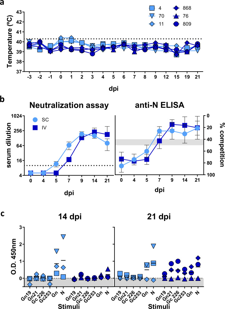
In the present study, we evaluated the immunogenicity, safety and protective efficacy of the attenuated RVFV-40Fp8 strain in natural hosts (non-pregnant ewes) and in a highly susceptible host infection model such as pregnant ewes in the first third of pregnancy. Our results confirm the immunogenicity of 40Fp8 administration in non-pregnant and pregnant ewes, as well as the absence of foetal damage even after a high-dose vaccination regime in pregnant ewes. In addition, the ewes and their foetuses were protected against a virulent RVFV-56/74 strain challenge, as shown by comparative evaluation of blood, serum and different tissue samples from vaccinated and non-vaccinated pregnant ewes. These results confirm the potential use of 40Fp8 as a RVF live-attenuated vaccine candidate and pave the way for further clinical developments.


Conflict of interest statement

Competing interests: A.B., B.B. and S.M. are the inventors of patent WO/2021/245313 (application PCT/ES2021/070403), which was filed with the Spanish National Research Council (Agencia Estatal Consejo Superior de Investigaciones Científicas, CSIC) and is currently being extended to several third countries. This manuscript supports the safety and efficacy of a vaccine candidate that is the subject of patent protection. All other authors declare no competing interests.

Figures

Fig. 1
Fig. 1. Experimental procedures in sheep.
Analysis of immunogenicity (a, b), safety (c) or protective efficacy (d) of the 40Fp8 vaccine strain in pregnant animals. Sheep identification numbers (#) are indicated. GD gestational day, IV intravenous inoculation, SC subcutaneous inoculation, dpi days post-immunization, dpc days post challenge.
Fig. 2
Fig. 2. Comparative evaluation of rectal temperatures, viremia levels, liver enzymes and RVFV-specific antibodies.
a Sheep rectal temperature records before and after virus inoculation. The horizontal black dotted line marks hyperthermia. b Viremia levels obtained upon titration in cell culture. The values displayed correspond to 50% tissue culture infectious dose per mL (TCID50/mL). Sensitivity limit of the assay is indicated by a horizontal dotted line. For the purposes of this graph, negative samples were assigned an arbitrary value of 100. c Kinetics of hepatic enzymes plasmatic levels after virus inoculation. AST Aspartate transaminase; GGT Gamma-glutamyltransferase and LDH Lactate dehydrogenase. The shaded areas depict normal range values. Values given in International Units (IU) per mL. d Kinetics of serum antibody responses. Each symbol represents values for individual sheep at the days of sacrifice (4, 7, 8), identified as in (a). Left panel: neutralizing antibodies. The values correspond to the reciprocal of serum dilution causing a 50% reduction of infectivity in cell culture. Right panel: anti-nucleoprotein antibodies. Values correspond to competition percentages as determined by the IDVet ELISA test. Shaded area between 40 and 50% indicates the doubtful status as defined by the provider. Red symbols: sheep inoculated with the wild-type 56/74 strain (n = 3). Blue symbols: sheep inoculated with 40Fp8 virus (n = 2). Individual identification of the 5 animals are indicated in (a).
Fig. 3
Fig. 3. Comparison of intravenous (IV) or subcutaneous (SC) 40Fp8 delivery.
a Evolution of rectal temperature in ewes after SC (light blue) or IV (dark blue) inoculation with 40Fp8 virus. Identification numbers of the 6 animals, 3 per group, are indicated. The horizontal black dotted line marks hyperthermia. b Serological determination of neutralizing antibody titers by TCID50 microneutralization assay (left panel) and competition ELISA anti-N (right panel). Both graphs show mean plus SD values. The dotted line depicts the sensitivity threshold (1/10 dilution). The gray shaded area depicts the doubtful status as defined by the IDVet ELISA test. c Detection of IFNγ in plasma by capture ELISA. Blood samples taken at 14 or 21 days after SC or IV 40Fp8 inoculation were re-stimulated with recombinant RVFV Gn or N proteins or peptides derived from the Gn (#19 and #21) or Gc (#226 and #253) protein sequences. Each symbol denotes individual values (after subtraction of values derived from unstimulated plasma) and horizontal bars denote mean values. Sheep identification as in (a).
Fig. 4
Fig. 4. Effect of 40Fp8 inoculation in pregnant ewes.
a Rectal temperature kinetics (mean ± SD) in ewes inoculated subcutaneously with 107 pfu of the 40Fp8 virus (n = 12) and in mock-inoculated ewes (n = 2). The horizontal dotted line marks hyperthermia. b Evaluation of foetal development. Mean and individual values for size and weight of foetuses found after necropsy at different days of gestation (GD). Number of foetuses for each gestation day is shown at Supplementary Table 2/GD 60: n = 7; GD 63: n = 9; GD 70: n = 8; GD 86: n = 3; mock samples: n = 3. Blue symbols, vaccinated; gray symbols, mock control (*). c Macroscopic images showing representative examples of the livers, foetuses and placentas of immunized ewes and non-immunised control ewes at 30 days post-immunisation (dpi)/86 gestational days (GD). Note the absence of the scattered necrotic lesions, characteristic of infected RVFV animals, the normal appearance of the placentas, and the presence of live foetuses with equivalent developmental parameters.
Fig. 5
Fig. 5. Molecular detection of 40Fp8 after inoculation in pregnant ewes (n = 12).
a Detection of viral RNA by RT-qPCR in blood samples taken from umbilical (foetus) or jugular veins of pregnant ewes at various days after inoculation with the 40Fp8 virus. b Detection of viral RNA by RT-qPCR in foetal and/or maternal tissues at various times after inoculation with the 40Fp8 virus. Data from liver, spleen and placentome (cotyledon) collected from pregnant ewes as well as data from liver, spleen and brain taken from foetuses are shown. Foetal spleen samples from 7 to 14 dpi were not retrieved. Blue symbols, vaccinated; gray symbols, mock control (*). The shaded areas indicate the sensitivity limit of the assay. The dotted lines indicate the relation between the Cq obtained and the corresponding viral infectious load or pfu equivalents.
Fig. 6
Fig. 6. Humoral responses upon 40Fp8 inoculation in pregnant ewes (n = 12).
Kinetics of neutralizing antibody levels assessed by microneutralization assay in sheep sera obtained on the indicated dates after vaccination with 40Fp8 virus. The dotted line indicates the sensitivity limit of the assay (left panel). Kinetics of induction of anti-nucleoprotein N antibodies assessed by competitive ELISA (IDVet RVFV screen test). The shaded area indicates the doubtful status as defined by the provider (right panel). Median and interquartile range as well as individual values are represented. Blue symbols, vaccinated; gray symbols, mock control (*).
Fig. 7
Fig. 7. Analysis of protection after RVFV virulent challenge in 40Fp8 vaccinated pregnant ewes.
a Rectal temperatures of individual ewes before and after virus challenge (red vertically dotted line). The horizontal black dotted line marks hyperthermia. b Macroscopic lesions and scores observed in sheep upon 56/74 challenge. When present, lesions were scored according to their extent in the tissues and the severity of the lesion itself between 1 (focal/minimal lesions) and 4 (diffuse/severe lesions). c Representative images of liver, foetuses and placentas found in immunized (upper images) and non-immunized sheep (bottom) after challenge. Scattered foci of necrotic lesions were found in the livers of non-immunized control ewes (white arrows) along with the presence of foetuses found dead and collected before imminent abortion. In contrast, livers without lesions and live foetuses were found in the 40Fp8 immunized ewes. Acute purulent metritis (yellow arrow) was observed in a non-immunized control ewe that aborted at 17 dpc, in contrast to the normal aspect placenta of a protected immunized ewe that was euthanized at 21 dpc.
Fig. 8
Fig. 8. Pathologic findings in non-vaccinated sheep and foetal tissues.
Representative images of histopathological lesions (haematoxylin-eosin staining) and immunohistochemical detection of RVFV antigen in different tissues obtained from pregnant ewes not immunized with the 40Fp8 strain and their foetuses at different days after challenge. Note the presence of hepatocytes immunolabelled against viral antigen within necrotic foci in the liver of non-immunized ewes at 4 and 7 dpc (a, e, 40x). Viral antigen was also occasionally observed in the placentomes at 4 dpc (b, 10x; c, d, 40x), specifically in maternal epithelial cells (red arrows). However, at 7 dpc, antigen-positive cells were visible throughout the placentomes of non-immunized ewe #6, both in the maternal epithelial cells/syncytial cells (red arrows) and in the foetal trophoblasts (black arrows) (f, 10x; g, 40x). Viral antigen was detected in brain, kidney and liver of foetuses before imminent abortion sampled at 7 dpc from ewe #6, but not in foetuses sampled at 4 dpc (h-m, 40x). Caruncular endometrium of ewe #4 aborted on 17 dpc revealed the presence of areas of coagulative necrosis and haemorrhages (asterisk), foci of mineralization (black arrowhead) and lymphoplasmacytic and neutrophilic infiltrates (black arrow) (n, 10x; o, magnified area, 40x). Remains of placenta from the aborted foetus of ewe #4 (p, 10x; q, magnified area, 40x). Note the presence of lymphoplasmacytic and neutrophilic infiltrates (black arrow) as well as the presence of viral antigen in the placental epithelial cells (black arrowheads). (f) foetal villus; (m); maternal villus; (mct): maternal connective tissue.
Fig. 9
Fig. 9. Molecular analysis after virus challenge.
a RT-qPCR analysis of viral RNA in vaccinated ewes and foetal (umbilical vein) blood before and after challenge. b Detection of viral RNA in sheep (liver and placentome) or foetal (liver, spleen, brain) tissue samples by RT-qPCR. In a and b, the shaded area indicates the sensitivity limit of the assay. Red daggers indicate that no foetuses were found in control sheep #4 (aborted on 17 dpc, and euthanized). Blue symbols, 40Fp8-vaccinated; red symbols, mock-vaccinated.
Fig. 10
Fig. 10. Humoral responses in vaccinated and challenged ewes.
Neutralizing anti-RVFV antibodies (nAbs) detected in sera by virus microneutralization assay in cell culture. Blue symbols, 40Fp8-vaccinated; red symbols, mock-vaccinated. The dotted line indicates sensitivity threshold (left panel). Detection of serum anti-nucleoprotein antibodies by competitive ELISA (IDVet RVF screen Kit). The shaded area indicates the doubtful status as defined by the provider (right panel) Median and interquartile range (for n > 3) as well as individual values are represented.

Similar articles

References

    1. Easterday, B. C. Rift valley fever. Adv. Vet. Sci.10, 65–127 (1965). - PubMed
    1. El Mamy, A. B. et al. Unexpected Rift Valley fever outbreak, northern Mauritania. Emerg. Infect. Dis.17, 1894–1896 (2011). - PMC - PubMed
    1. El Mamy, A. B. et al. Comprehensive phylogenetic reconstructions of Rift Valley fever virus: the 2010 northern Mauritania outbreak in the Camelus dromedarius species. Vector Borne Zoonotic Dis.14, 856–861 (2014). - PMC - PubMed
    1. Turell, M. J. & Bailey, C. L. Transmission studies in mosquitoes (Diptera: Culicidae) with disseminated Rift Valley fever virus infections. J. Med. Entomol.24, 11–18 (1987). - PubMed
    1. Gardela, J. et al. Tissue distribution and transmission of Rift Valley fever phlebovirus in European Culex pipiens and Aedes albopictus mosquitoes following intrathoracic inoculation. J. Gen. Virol. 2024;105. Epub 2024/09/21. 10.1099/jgv.0.002025. - PMC - PubMed

LinkOut - more resources