. 2025 Aug 29;41(1):128.
doi: 10.1007/s10565-025-10069-9.

DNMT1 recruits RUNX1 and represses FOXO1 transcription to inhibit anti-inflammatory activity of regulatory T cells and augments sepsis-induced lung injury

Affiliations

DNMT1 recruits RUNX1 and represses FOXO1 transcription to inhibit anti-inflammatory activity of regulatory T cells and augments sepsis-induced lung injury

Jurong Ding et al. Cell Biol Toxicol. .

Abstract

Sepsis-induced lung injury (ALI) is a critical condition characterized by excessive immune responses and tissue damage. Previous evidence has underscored an upregulation pattern of DNA methyltransferase 1 (DNMT1) in sepsis. This study reveals the key role of DNMT1 in modulating regulatory T cell (Treg) activity in septic ALI. A septic mouse model was generated through cecal ligation and puncture. Treatment with either DNMT1 antagonist Thioguanine (ThG) or AAV-sh-DNMT1 significantly reduced immune cell infiltration, reduced production of pro-inflammatory cytokines, and increasing production of anti-inflammatory cytokines in the bronchoalveolar lavage fluid (BALF) of mice, alongside improved lung pathology and integrity. Furthermore, the DNMT1 inhibition or silencing significantly enhanced population of FOXP3+ Tregs in the BALF and lung tissue. Similar trends were observed in mice with specific DNMT1 deletion in CD4+ T cells (DNMT1-CD4-ko). Regarding the mechanism, we observed that DNMT1 represses transcription of forkhead box O1 (FOXO1) by recruiting RUNX family transcription factor 1 (RUNX1) to the FOXO1 promoter. FOXO1-specific knockout in CD4+ T cells reduced anti-inflammatory activity of Tregs. Additionally, administration of the CD25 antibody exacerbated sepsis-induced ALI in DNMT1-CD4-ko mice. Collectively, these findings illustrate that targeting DNMT1 interacts with RUNX1 to repress transcription of FOXO1, which reduces immunomodulatory activity of Tregs and augments inflammatory cascades in septic lung injury.

Keywords: DNMT1; FOXO1; Lung injury; RUNX1; Sepsis; Tregs.


Conflict of interest statement

Declarations. Ethics approval and consent to participate: This study was approved by the Shanghai Pulmonary Hospital Ethics Committee. All animal experiments complied with the guidelines of the Shanghai Pulmonary Hospital Animal Care and Use Committee (Approval number: K21-354). Consent for publication: Not applicable. Competing interests: The authors declare no competing interests.

Figures

Fig. 1
Fig. 1
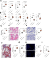
DNMT1 inhibition attenuates sepsis-induced ALI in mice. A Schematic presentation of the septic mouse model induced via CLP challenge, followed by intratracheal injection of either DNMT1 antagonist ThG (3 mg/kg/day) or AAV9-encapsulated sh-DNMT1. B Number of macrophages, lymphocytes, neutrophils, and eosinophils in mouse BALF. C ELISA analysis of TNF-α, IL-6, IL-1β, MCP-1, IL-10 and TGF-β1 concentrations in mouse BALF. D Evans Blue staining of vascular permeability in mouse lung tissue. E HE staining to analyze pathological injury in mouse lung tissues. F PAS staining to assess glycoprotein deposition in mouse lung tissues. G Flow cytometric analysis of CD4+CD25+FOXP3+ T cells in mouse BALF. H Immunofluorescence detection of FOXP3+ cells in mouse lung tissue (Red: FOXP3+; Blue: DAPI). Each group contains six mice, and three independent replications were conducted for each sample Differences between groups were compared by one-way ANOVA followed by Tukey’s post-hoc tests. p < 0.05 was considered statistically significant
Fig. 2
Fig. 2
DNMT1 deficiency in CD4+ T cells alleviates sepsis-induced AKI in mice by enhancing the immunosuppressive function of Tregs. DNMT1-CD4-Cre mice with specific DNMT1 deletion in CD4+ T cells (DNMT1-CD4-ko) were generated, followed by CLP challenge to induce sepsis. CLP-challenged DNMT1fl/fl mice were set to controls. A Number of macrophages, neutrophils, eosinophils, and lymphocytes in mouse BALF. B ELISA analysis of TNF-α, IL-6, IL-1β, MCP-1, IL-10 and TGF-β1 concentrations in mouse BALF. C Evans Blue staining of vascular permeability in mouse lung tissue. D HE staining to analyze pathological injury in mouse lung tissues. E PAS staining to assess glycoprotein deposition in mouse lung tissues. F Masson's trichrome staining to detect collagen deposition in lung tissues. G qPCR analysis of mRNA levels of fibrosis-related genes (FN1, SPP1, and COL1A1) in mouse lung tissues. H Flow cytometry analysis of CD4+CD25+FOXP3+ T cells in mouse BALF. I Immunofluorescence detection of FOXP3+ cells in mouse lung tissue (Red: FOXP3+; Blue: DAPI). J Flow cytometric analysis of CD4+CD25+CTLA4+ T cells in mouse BALF. Each group contains six mice, and three independent replications were conducted for each sample. Differences between groups were compared by unpaired t tests, or by the two-way ANOVA followed by Tukey’s post-hoc tests. p < 0.05 was considered statistically significant
Fig. 3
Fig. 3
DNMT1 represses FOXO1 expression. A Naive CD4+ T cells were isolated from DNMT1fl/fl or DNMT1-CD4-ko mice. These cells were the stimulated with These cells were stimulated with anti-CD3 and anti-CD28 antibodies, followed by induction with TGF-β1 and IL-2 to promote their differentiation into Tregs. B Volcano plots for DEGs in Tregs from DNMT1fl/fl and DNMT1-CD4-ko mice identified through RNA-seq analysis. C~D qPCR and WB analysis of FOXO1 levels in Tregs. E CpG island upstream of the FOXO1 promoter predicted using the TUBIC software. F BSP-qPCR detection of the CpG island in FOXO1 promoter in Tregs. G Binding of DNMT1 to the FOXO1 promoter determined using ChIP-qPCR assays. H Regulation of DNMT1 on the transcription activity of FOXO1 determined using luciferase reporter gene assays. Six independent experiments were performed. Differences between groups were compared by unpaired t tests, or by the two-way ANOVA followed by Tukey’s post-hoc tests. p < 0.05 was considered statistically significant
Fig. 4
Fig. 4
DNMT1 recruits RUNX1 to repress FOXO1 transcription. A Volcano plots of DNMT1-interacting proteins identified by IP-MS. B Co-localization of DNMT1 and RUNX1 in extracted Tregs or mouse lung tissue sections determined using double-label fluorescence staining assays (Red: RUNX1+; Green: DNMT1+; Blue: DAPI). C Immunofluorescence detection of RUNX1 nuclear localization in Tregs (Red: RUNX1+; Green: DNMT1+; Blue: DAPI). D The interaction between RUNX1 and DNMT1 determined using Co-IP and WB assays. E Binding of RUNX1 to the FOXO1 promoter determined using ChIP-qPCR assays. F Regulation of RUNX1 on the transcription activity of FOXO1 determined using luciferase reporter gene assays. G-H qPCR and WB analysis of RUNX1 and FOXO1 levels in 293 T cells after RUNX1 overexpression. Six independent experiments were performed. Differences between groups were compared by unpaired t tests, or by the two-way ANOVA followed by Tukey’s post-hoc tests. p < 0.05 was considered statistically significant
Fig. 5
Fig. 5
FOXO1 deficiency in CD4+ T cells reduces anti-inflammatory activity of Tregs. FOXO1-CD4-ko mice were generated, followed by CLP challenge to induce sepsis. CLP-challenged DNMT1fl/fl mice were set to controls. A Number of macrophages, neutrophils, eosinophils, and lymphocytes in BALF. B ELISA analysis of inflammatory cytokines NF-α, IL-6, IL-1β, MCP-1, IL-10 and TGF-β1 concentrations in mouse BALF. C Evans Blue staining of vascular permeability in mouse lung tissue. D HE staining to analyze pathological injury in mouse lung tissues. E PAS staining to assess glycoprotein deposition in mouse lung tissues. F Masson's trichrome staining to detect collagen deposition in lung tissues. G Flow cytometry analysis of CD4+CD25+FOXP3+ T cells in mouse BALF. H Immunofluorescence detection of FOXP3+ cells in mouse lung tissues (Red: FOXP3+; Blue: DAPI). J Flow cytometric analysis of CD4+CD25+CTLA4+ T cells in mouse BALF. Each group contains six mice, and three independent replications were conducted for each sample. Differences between groups were compared by unpaired t tests. p < 0.05 was considered statistically significant
Fig. 6
Fig. 6
ThG also alleviates septic lung injury in FOXO1-CD4-ko mice. A Number of macrophages, neutrophils, eosinophils, and lymphocytes in BALF. B ELISA analysis of inflammatory cytokines in BALF. C Evans Blue staining of vascular permeability in mouse lung tissue. D HE staining to analyze pathological injury in mouse lung tissues. E PAS staining to assess glycoprotein deposition in mouse lung tissues. F Flow cytometry analysis of CD4+CD25+FOXP3+ T cells in mouse BALF. G Immunofluorescence detection of FOXP3+ cells in mouse lung tissues (Red: FOXP3+; Blue: DAPI). Each group contains six mice, and three independent replications were conducted for each sample. Differences between groups were compared by unpaired t tests. p < 0.05 was considered statistically significant
Fig. 7
Fig. 7
CD25 antibody exacerbates sepsis-induced ALI in DNMT1-CD4-ko mice. DNMT1-CD4-ko mice were treated with anti-CD25 to deplete Tregs, followed by sepsis modeling. A Flow cytometry analysis of CD4+CD25+FOXP3+ T cells in mouse BALF. B Immunofluorescence detection of FOXP3+ cells in mouse lung tissues (Red: FOXP3+; Blue: DAPI). C Number of macrophages, neutrophils, eosinophils, and lymphocytes in BALF. D ELISA analysis of inflammatory cytokines in BALF. E Evans Blue staining of vascular permeability in mouse lung tissue. F HE staining to analyze pathological injury in mouse lung tissues. G PAS staining to assess glycoprotein deposition in mouse lung tissues. H Masson's trichrome staining to detect collagen deposition in lung tissues. Each group contains six mice, and three independent replications were conducted for each sample. Differences between groups were compared by unpaired t tests. p < 0.05 was considered statistically significant
Fig. 8
Fig. 8
Graphic Abstract. DNMT1 as an essential regulator in sepsis-induced ALI by repressing FOXO1 and impairing Treg function. Inhibiting DNMT1 restores FOXO1 expression and enhances Treg-mediated immune suppression, leading to reduced inflammation and improved lung integrity

Similar articles

References

    1. Allos H, Al Dulaijan BS, Choi J, Azzi J. Regulatory T Cells for More Targeted Immunosuppressive Therapies. Clin Lab Med. 2019;39(1):1–13. 10.1016/j.cll.2018.11.001. - PMC - PubMed
    1. Barutcu AR, Hong D, Lajoie BR, McCord RP, van Wijnen AJ, Lian JB, et al. RUNX1 contributes to higher-order chromatin organization and gene regulation in breast cancer cells. Biochim Biophys Acta. 2016;1859(11):1389–97. 10.1016/j.bbagrm.2016.08.003. - PMC - PubMed
    1. Carrette F, Fabre S, Bismuth G. FOXO1, T-cell trafficking and immune responses. Adv Exp Med Biol. 2009;665:3–16. 10.1007/978-1-4419-1599-3_1. - PubMed
    1. Chan JD, Scheffler CM, Munoz I, Sek K, Lee JN, Huang YK, et al. FOXO1 enhances CAR T cell stemness, metabolic fitness and efficacy. Nature. 2024;629(8010):201–10. 10.1038/s41586-024-07242-1. - PMC - PubMed
    1. Chao Y, Huang W, Xu Z, Li P, Gu S. Effect of RUNX1/FOXP3 axis on apoptosis of T and B lymphocytes and immunosuppression in sepsis. Open Med (Wars). 2023;18(1):20230728. 10.1515/med-2023-0728. - PMC - PubMed

MeSH terms