. 2025 Aug 29;15(1):31918.
doi: 10.1038/s41598-025-17961-8.

Nanofat promotes wound healing in skin following exposure to ionizing radiation

Affiliations

Nanofat promotes wound healing in skin following exposure to ionizing radiation

Ettore Limido et al. Sci Rep. .

Abstract

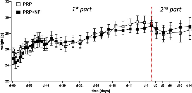
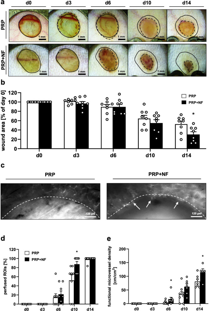
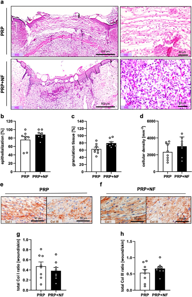
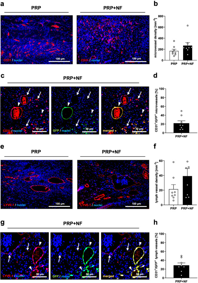
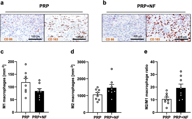
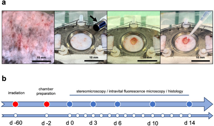
Radiotherapy, while effective in cancer treatment, can lead to side effects, such as radiodermatitis with potential long-term consequences including telangiectasias, ulceration and fibrosis of the skin, eventually resulting in impaired wound healing. In this study, we analyzed whether the healing of such challenging wounds can be improved by nanofat (NF). NF is generated by mechanical emulsification and filtration of fat samples and, thus, is a random mixture of adipose-derived stem cells, microvascular fragments, extracellular matrix components and growth factors. Two months after localized ionizing radiation of the skin with a total dose of 20 Gy, full-thickness wounds were created in dorsal skinfold chambers of mice, which were filled with platelet-rich plasma (PRP; control, n = 8) or NF fixed in PRP (PRP + NF, n = 8). The healing process was assessed by means of stereomicroscopy, intravital fluorescence microscopy, histology and immunohistochemistry over 14 days. The closure of PRP + NF-treated wounds was accelerated, as indicated by significantly smaller wound areas on day 14 when compared to controls. This was associated with a higher density of blood-perfused microvessels inside the wounds. Moreover, PRP + NF-treated wounds showed a tendency towards an improved granulation tissue formation, lymphatic drainage and M2/M1 macrophage ratio. Taken together, these findings suggest that the application of NF represents a promising therapeutic strategy for the management of complex wounds in irradiated skin.

Keywords: Dorsal skinfold chamber; Nanofat; Platelet-rich plasma; Radiotherapy; Vascularization; Wound healing.


Conflict of interest statement

Declarations. Competing interests: The authors declare no competing interests.

Figures

Fig. 1
Fig. 1
Development of body weight. Body weight (g) of mice that underwent localized ionizing radiation of the dorsal skinfold on day − 60 and were equipped with dorsal skinfold chambers on day − 2 (red dotted line). Within the chambers, wounds were created on day 0, which were then treated with PRP (white squares; n = 8) or a combination of PRP and NF (PRP + NF; black squares; n = 8). Mean ± SEM.
Fig. 2
Fig. 2
In vivo microscopy of healing wounds within dorsal skinfold chambers. (a) Stereomicroscopic images of a PRP-treated and a PRP + NF-treated wound on days 0, 3, 6, 10 and 14 (solid lines = initial wound borders; broken lines = wound borders at the indicated time points). (b) Wound area (% of day 0) of PRP-treated (white bars; n = 8) and PRP + NF-treated (black bars; n = 8) wounds on days 0, 3, 6, 10 and 14, as assessed by stereomicroscopy. Mean ± SEM. *p < 0.05 vs. PRP. (c) Intravital fluorescence microscopic images of a PRP-treated and a PRP + NF-treated wound with first blood-perfused microvessels at the wound border (arrows) on day 6 (broken lines = wound borders). (d, e) Perfused ROIs (d, %) and functional microvessel density (e, cm/cm²) of PRP-treated (white bars; n = 8) and PRP + NF-treated (black bars; n = 8) wounds on days 0, 3, 6, 10 and 14, as assessed by intravital fluorescence microscopy. Mean ± SEM. *p < 0.05 vs. PRP.
Fig. 3
Fig. 3
Tissue formation in healing wounds. (a) HE-stained sections (left panels = overview of the wounds showing their epithelialization; right panels = higher magnification of the granulation tissue in the wound center) of a PRP-treated and a PRP + NF-treated wound on day 14. (b-d) Epithelialization (b, %), granulation tissue formation (c, %) and cellular density (d, cells/mm− 2) of PRP-treated (white bars; n = 8) and PRP + NF-treated (black bars; n = 8) wounds on day 14, as assessed by histology. Mean ± SEM. (e, f) Immunohistochemical detection of Col I and Col III in a PRP-treated and a PRP + NF-treated wound on day 14. (g, h) Total Col I (g) and Col III (h) ratio (wound/normal skin) of PRP-treated (white bars; n = 8) and PRP + NF-treated (black bars; n = 8) wounds on day 14, as assessed by immunohistochemistry. Mean ± SEM.
Fig. 4
Fig. 4
Vascularization and lymphatic drainage of healing wounds. (a) Immunohistochemical detection of CD31+ microvessels in a PRP-treated and a PRP + NF-treated wound on day 14. (b) Microvessel density (mm− 2) of PRP-treated (white bars; n = 8) and PRP + NF-treated (black bars; n = 8) wounds on day 14, as assessed by immunohistochemistry. Mean ± SEM. (c) Immunohistochemical detection of CD31+/GFP (arrows) and CD31+/GFP+ (arrowheads) microvessels in a PRP + NF-treated wound on day 14. (d) CD31+/GFP+ microvessels (%) in PRP + NF-treated wounds (black bar; n = 8) on day 14, as assessed by immunohistochemistry. Mean ± SEM. (e) Immunohistochemical detection of LYVE-1+ lymph vessels in a PRP-treated and a PRP + NF-treated wound on day 14. (f) Lymph vessel density (mm− 2) of PRP-treated (white bars; n = 8) and PRP + NF-treated (black bars; n = 8) wounds on day 14, as assessed by immunohistochemistry. Mean ± SEM. (g) Immunohistochemical detection of LYVE-1+/GFP (arrows) and LYVE-1+/GFP+ (arrowheads) lymph vessels in a PRP + NF-treated wound on day 14. (H) LYVE-1+/GFP+ lymph vessels (%) in PRP + NF-treated wounds (black bar; n = 8) on day 14, as assessed by immunohistochemistry. Mean ± SEM.
Fig. 5
Fig. 5
Macrophage infiltration of healing wounds. (a, b) Immunohistochemical detection of CD86+ M1 and CD163+ M2 macrophages in a PRP-treated and a PRP + NF-treated wound on day 14. (c-e) M1 macrophages (c, mm− 2), M2 macrophages (d, mm− 2), and M2/M1 macrophage ratio (e) in PRP-treated (white bars; n = 8) and PRP + NF-treated (black bars; n = 8) wounds on day 14, as assessed by immunohistochemistry. Mean ± SEM.
Fig. 6
Fig. 6
Experimental setting of the present study. (a) Left panel: Signs of radiodermatitis in the skin on the back of a mouse 3 weeks after localized ionizing radiation with a total dose of 20 Gy. Middle-left panel: Observation window of the dorsal skinfold chamber immediately before wound creation with a 4-mm biopsy punch (arrow). Middle-right panel: Observation window immediately after wound creation. Right panel: Filling of the wound with a combination of PRP and NF by means of a pipette. (b) Timeline of the in vivo experiments according to the detailed description in the materials and method section.

References

    1. Jensen, J. M. & Proksch, E. The skin’s barrier. G Ital. Dermatol. Venereol.144, 689–700 (2009). - PubMed
    1. Wang, P. H., Huang, B. S., Horng, H. C., Yeh, C. C. & Chen, Y. J. Wound healing. J. Chin. Med. Assoc.81, 94–101 (2018). - PubMed
    1. Fabbrocini, G. et al. Chemotherapy and skin reactions. J. Exp. Clin. Cancer Res.31, 50 (2012). - PMC - PubMed
    1. Russi, E. G. et al. Acute skin toxicity management in head and neck cancer patients treated with radiotherapy and chemotherapy or EGFR inhibitors: literature review and consensus. Crit. Rev. Oncol. Hematol.96, 167–182 (2015). - PubMed
    1. Sønstevold, T., Johannessen, A. C. & Stuhr, L. A rat model of radiation injury in the mandibular area. Radiat. Oncol.10, 129 (2015). - PMC - PubMed

LinkOut - more resources