. 2025 Aug 30;25(1):566.
doi: 10.1186/s12866-025-04215-w.

Role of P38 lipoprotein in Mycoplasma penetrans adhesion to human urothelial cells

Affiliations

Role of P38 lipoprotein in Mycoplasma penetrans adhesion to human urothelial cells

Kaihua Zhang et al. BMC Microbiol. .

Abstract

Mycoplasma penetrans, a bacterium detected in individuals seropositive for HIV and phylogenetically clustered with M. muris, may contribute to the progression of Acquired Immune Deficiency Syndrome (AIDS). Cellular adhesion is essential for Mycoplasma infection of host cells. M. penetrans exhibits the capacity to adhere to and invade human cells, precipitating diseases of the genital and urinary tracts. However, the proteinaceous mediators of its adhesion remain largely elusive. The P35 family lipoprotein, encoded by the mpl gene, is a prominent surface lipoprotein of M. penetrans. Here, we investigated the role of P38, a member of the P35 family, in the adhesion of M. penetrans to human urothelial cells (SV-HUC-1). We expressed and purified recombinant P38 (rP38) and confirmed its localization using Western blot. Adhesion assays, adhesion inhibition assays, and adhesion competition assays were performed to evaluate the adhesive properties of rP38 and M. penetrans. Our findings indicated that P38 localizes to the cell membrane of M. penetrans. Both rP38 and M. penetrans adhered to SV-HUC-1 cells, with optimal adhesion observed at 60 μg/mL for rP38 and 1 × 10⁷ CCU (Colony-Changing Units)/mL for M. penetrans. Anti-rP38 serum partially inhibited M. penetrans adhesion to SV-HUC-1 cells, and rP38 competed with M. penetrans for binding to SV-HUC-1 cells. These results suggest that P38 may function as an adhesin of M. penetrans, providing insights into its pathogenic mechanisms.

Keywords: Mycoplasma; Mycoplasma penetrans; Adhesion protein; Lipoprotein; P38.


Conflict of interest statement

Declarations. Ethics approval and consent to participate: The antibody preparation using animals was approved by the Ethics Committee of the University of South China, and all animal procedures were conducted in accordance with the institutional regulations (No. 2023–38). Consent for publication: Not applicable. Competing interests: The authors declare no competing interests.

Figures

Fig. 1
Fig. 1
Expression and Purification of the P38 Protein. A Restriction endonuclease analysis of the pET-28a/P38 prokaryotic expression plasmid. Lane M: KB Ladder. Lane 1: pET-28a/P38 plasmid digested with BamHI and XhoI. Lane 2: Undigested pET-28a/P38 plasmid. B SDS-PAGE analysis of recombinant P38 protein expression and purification. Lane 1: Total protein from E. coli BL21(DE3) cells cultured at 37 °C without IPTG induction. Lanes 2 and 3: Supernatant protein from lysed E. coli BL21(DE3) cells harboring the pET-28a/P38 plasmid and induced with 0.6 mM IPTG at 37 °C. Lane 4: Precipitated protein from lysed E. coli BL21(DE3) cells harboring the pET-28a/P38 plasmid and induced with 0.6 mM IPTG at 37 °C. Lanes 5–9: Purified recombinant P38 protein eluted with 60, 80-, 90-, 100-, and 150-mM imidazole solution, respectively. C Western blot analysis of purified rP38 using an anti-His antibody. Lane 1: Supernatant protein from lysed E. coli BL21(DE3) cells without the pET-28a/P38 plasmid. Lane 2: Supernatant protein from lysed E. coli BL21(DE3) cells harboring the pET-28a/P38 plasmid and induced with 0.6 mM IPTG. Lane 3: Supernatant protein from lysed E. coli BL21(DE3) cells harboring the pET-28a/P38 plasmid without IPTG induction. The molecular weight of P38 is approximately 38.37 kDa
Fig. 2
Fig. 2
Localization of P38 protein in M. penetrans. A This panel shows the SDS-PAGE result of protein profiles of the different M. penetrans fractions. Lane M: Protein marker. Lane 1: Total M. penetrans cell lysate. This lane shows the complex mixture of all proteins present in the cells. Lane 2: Cytoplasmic protein fraction. Lane 3: Membrane protein fraction. B We used the anti-rP38 antibody to detect P38 protein specifically within the fractions shown in Panel A. The presence of a band at approximately38 kDa in lane 1 (Total protein) confirms the presence of P38 in the whole-cell lysate. Lane 2 (Cytoplasmic protein) shows a faint band, suggesting a small amount of P38 might be present in the cytoplasm, or possibly due to cross-contamination or non-specific binding of the antibody. Lane 3 (Membrane protein) shows a strong band, confirming the predominant localization of P38 in the membrane fraction
Fig. 3
Fig. 3
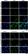
The adhesion of both rP38 and M. penetrans to SV-HUC-1 cells using immunofluorescence. Different concentrations of rP38 (20, 40, and 60 μg/mL) and M. penetrans (1 × 10⁶, 1 × 10⁷, and 1 × 10⁸ CCU/mL) were incubated with the cells. Detection was performed using anti-rP38 and anti-M. penetrans sera as primary antibodies. An Alexa Fluor 488-conjugated secondary antibody (green) targeted the primary antibodies, allowing visualization of the rP38 and M. penetrans bound to the SV-HUC-1 cells. Cell nuclei were counterstained with DAPI (blue). The intensity of the green fluorescence was measured by ImageJ. A representative result of each group is displayed. Scale bar: 20 μm
Fig.4
Fig.4
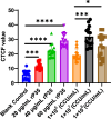
The average fluorescence analysis of adhesion assay results. ImageJ was employed to analyze the fluorescence density of adhesion assay. Fifteen cells were selected from the adhesion inhibition assay results for average fluorescence analysis. GraphPad Prism software was used for graphing. The data were presented with average fluorescence mean with SD. Significant difference analysis was performed using GraphPad Prism ordinary one-way ANOVA. *: P < 0.05; *** P: < 0.001, ****: P < 0.0001
Fig. 5
Fig. 5
Adhesion Inhibition Assay of the rP38 and M. penetrans to SV-HUC-1 cells. Anti-rP38 serum were used to inhibit adhesive process of P38 protein and M. penetrans. These assays were followed by visualization with Alexa Fluor 488-conjugated Affineur goat anti-rabbit IgG (green). The cell nuclei were stained with DAPI (blue). (Scale bar: 20 μm.)
Fig. 6
Fig. 6
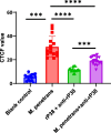
Analysis of the average fluorescence intensity in adhesion inhibition assay. The fluorescence intensity was analyzed using ImageJ software. Fifteen cells were selected from the adhesion inhibition assay for average fluorescence analysis. GraphPad Prism software was used for graphing. The data were presented with average fluorescence mean with SD. Significant difference analysis was performed using GraphPad Prism ordinary one-way ANOVA. *** P < 0.001, ****: P < 0.0001
Fig. 7
Fig. 7
Adhesion competition assay of recombinant P38 and M. penetrans to SV-HUC-1 cells. Fluorescence microscopy observation result. The primary antibody used was anti-M. penetrans serum. The SV-HUC-1 cells incubated with M. penetrans directly as a positive control. (Scale bar: 20 μm)
Fig. 8
Fig. 8
Analysis of the average fluorescence intensity in Adhesion competition assay. The fluorescence intensity was analyzed using ImageJ. Fifteen cells were selected from the adhesion adhesion competition assay for average fluorescence analysis. GraphPad Prism software was used for graphing. The data were presented with average fluorescence mean with SD. Significant difference analysis was performed using GraphPad Prism ordinary one-way ANOVA. **** P < 0.0001

Similar articles

References

    1. He J, et al. Insights into the pathogenesis of Mycoplasma pneumoniae. Mol Med Rep. 2016;14(5):4030–6. - PMC - PubMed
    1. Yu Y, et al. Mycoplasma hyopneumoniae evades complement activation by binding to factor H via elongation factor thermo unstable (EF-Tu). Virulence. 2020;11(1):1059–74. - PMC - PubMed
    1. Jambhekar A, Robin E, Le Boedec K. A systematic review and meta-analyses of the association between 4 mycoplasma species and lower respiratory tract disease in dogs. J Vet Intern Med. 2019;33(5):1880–91. - PMC - PubMed
    1. Luo H, et al. Mycoplasma pneumoniae lipids license TLR-4 for activation of NLRP3 inflammasome and autophagy to evoke a proinflammatory response. Clin Exp Immunol. 2021;203(1):66–79. - PMC - PubMed
    1. Li L, et al. Mycoplasma genitalium protein of adhesion induces inflammatory cytokines via cyclophilin A-CD147 activating the ERK-NF-κB pathway in human urothelial cells. Front Immunol. 2020;11:2052. - PMC - PubMed

MeSH terms

LinkOut - more resources