M1-BMDMs with Wnt5a deletion attenuate liver fibrosis by suppression of Wnt5a/Frizzled 2 axis in hepatic progenitors
- PMID: 40885967
- PMCID: PMC12398029
- DOI: 10.1186/s13578-025-01467-x
M1-BMDMs with Wnt5a deletion attenuate liver fibrosis by suppression of Wnt5a/Frizzled 2 axis in hepatic progenitors
Abstract
Background: Bone marrow-derived macrophages (BMDMs) regulate hepatic progenitor cells (HPCs) differentiation, potentially via the Wnt signaling pathway. While M1-polarized BMDMs (M1-BMDMs) exert anti-fibrotic effects in the liver, Wnt5a is implicated in fibrosis progression. The specific influence of Wnt5a levels within M1-BMDMs on HPCs fate and cirrhosis development remains unclear. This study aimed to elucidate the relationship between M1-BMDM-derived Wnt5a and HPCs differentiation during cirrhosis progression.
Methods: First, Wnt5a protein expression was assessed in liver biopsy tissues from patients with hepatitis B-associated liver fibrosis. Second, cirrhosis was induced in rats using CCl4/2-AAF. In week 9, rats received intravenous injections of M1-BMDMs with Wnt5a knockdown (M1-BMDM Wnt5a−KD) or overexpression (M1-BMDM Wnt5a−OE); peripheral BMDMs recruitment was blocked using a CCR2 inhibitor. Fibrosis progression, ductular reaction (DR), and HPC differentiation were evaluated. In vitro, WB-F344 cells subjected to frizzled 2 (Fzd2) knockdown (WB-F344 Fzd2−KD) or overexpression (WB-F344 Fzd2−OE) were cultured with conditioned medium from M1-BMDM Wnt5a−KD (CM Wnt5a−KD) or M1-BMDM Wnt5a−OE (CM Wnt5a−OE).
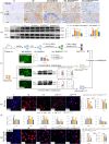
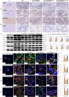
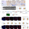
Results: In patients with hepatitis B-related fibrosis, hepatic Wnt5a expression increased progressively with METAVIR fibrosis grade. In the rat cirrhosis model, M1-BMDMs Wnt5a−KD attenuated fibrosis, whereas M1-BMDMs Wnt5a−OE exacerbated it. Mechanistically, in vivo injection of M1-BMDMs Wnt5a−KD significantly inhibited HPCs differentiation into biliary epithelial cells (BECs), while M1-BMDM Wnt5a−OE promoted this differentiation. In vitro, CM Wnt5a−KD inhibited the differentiation of WB-F344 cells into BECs; this inhibition was potentiated by Fzd2 knockdown in WB-F344 cells but abrogated by Fzd2 overexpression. Conversely, under CM Wnt5a−OE conditions, WB-F344 Fzd2−OE cells exhibited increased cholangiocytic differentiation, an effect largely negated by Fzd2 knockdown.
Conclusions: M1-BMDMs Wnt5a−KD demonstrated superior therapeutic efficacy against cirrhosis compared to unmodified M1-BMDMs. Wnt5a/Fzd2 signaling mediated the crosstalk between M1-BMDMs Wnt5a−KD and HPCs, revealing a novel therapeutic target for cirrhosis treatment.
Supplementary Information: The online version contains supplementary material available at 10.1186/s13578-025-01467-x.
Keywords: Bone marrow-derived macrophage; Ductular reaction; Hepatic progenitor cells; Liver cirrhosis; Wnt5a/Frizzled2 signaling axis.
Conflict of interest statement
Declarations. Ethics approval and consent to participate: Liver tissue slices from patients with hepatitis B-associated liver fibrosis were obtained from the biobank of the clinical study titled “Chinese Herbal Medicine Combined with Entecavir in Treating Hepatitis B Cirrhosis”. This clinical trial was approved by the Ethics Committee of Shuguang Hospital Affiliated to Shanghai University of TCM (Ethics approval No. 2014-331-27-01). The ethics of animal experimentation are as follows: (1) Title of the approved project: The effector mechanism of macrophage polarization in the regulation of hepatic progenitor cells differentiation and intervention in liver cirrhosis. (2) Name of the institutional approval committee or unit: Animal Research Committee at Shanghai University of TCM. (3) Ethics approval No. PZSHUTCM210611009. (4) Date of approval: June 11, 2021. Consent for publication: Not applicable. Competing interests: The authors declare that they have no competing interests.
Figures


Similar articles
-
[Research on the mechanism of gentiopicroside preventing macrophage-mediated liver fibrosis by regulating the MIF-SPP1 signaling pathway in hepatic stellate cells].Xi Bao Yu Fen Zi Mian Yi Xue Za Zhi. 2025 Jul;41(7):593-602. Xi Bao Yu Fen Zi Mian Yi Xue Za Zhi. 2025. PMID: 40620116 Chinese.
-
Secreted frizzled-related protein 5 (Sfrp5) decreases hepatic stellate cell activation and liver fibrosis.Liver Int. 2015 Aug;35(8):2017-26. doi: 10.1111/liv.12757. Epub 2015 Jan 20. Liver Int. 2015. PMID: 25488180
-
MBD2 deficiency attenuates CCl4-induced hepatic fibrosis by inhibiting M2 macrophage polarization.Int Immunopharmacol. 2025 Aug 6;164:115306. doi: 10.1016/j.intimp.2025.115306. Online ahead of print. Int Immunopharmacol. 2025. PMID: 40773897
-
Transient elastography for diagnosis of stages of hepatic fibrosis and cirrhosis in people with alcoholic liver disease.Cochrane Database Syst Rev. 2015 Jan 22;1(1):CD010542. doi: 10.1002/14651858.CD010542.pub2. Cochrane Database Syst Rev. 2015. PMID: 25612182 Free PMC article.
-
Granulocyte colony-stimulating factor with or without stem or progenitor cell or growth factors infusion for people with compensated or decompensated advanced chronic liver disease.Cochrane Database Syst Rev. 2023 Jun 6;6(6):CD013532. doi: 10.1002/14651858.CD013532.pub2. Cochrane Database Syst Rev. 2023. PMID: 37278488 Free PMC article.
References
-
- Ginès P, Castera L, Lammert F, Graupera I, Serra-Burriel M, Allen AM, et al. LiverScreen consortium investigators. Population screening for liver fibrosis: toward early diagnosis and intervention for chronic liver diseases. Hepatology. 2022;75(1):219–28. - PubMed
-
- Freeman RB Jr, Steffick DE, Guidinger MK, Farmer DG, Berg CL, Merion RM. Liver and intestine transplantation in the united states, 1997–2006. Am J Transpl. 2008;8(4 Pt 2):958–76. - PubMed
-
- Oakley F. Interrogating mechanisms of liver fibrosis with omics. Nat Rev Gastroenterol Hepatol. 2022;19(2):89–90. - PubMed
Grants and funding
LinkOut - more resources
Full Text Sources

