. 2025 Dec;47(1):2548613.
doi: 10.1080/0886022X.2025.2548613. Epub 2025 Aug 31.

Glycyrrhizin alleviates contrast-induced acute kidney injury via inhibiting HMGB1-mediated renal tubular epithelial cells ferroptosis

Affiliations

Glycyrrhizin alleviates contrast-induced acute kidney injury via inhibiting HMGB1-mediated renal tubular epithelial cells ferroptosis

Fang-Yuan Tian et al. Ren Fail. 2025 Dec.

Abstract

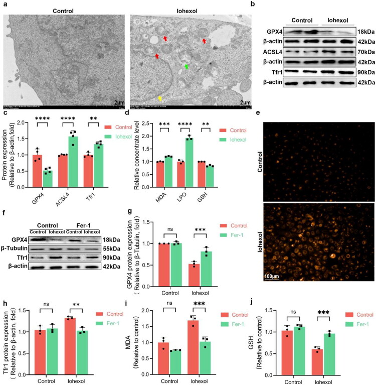
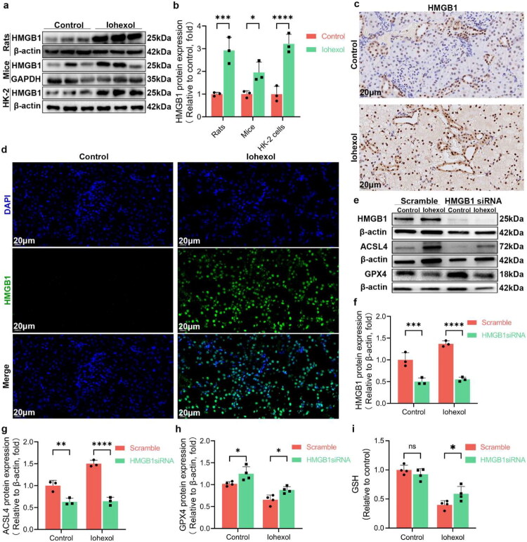
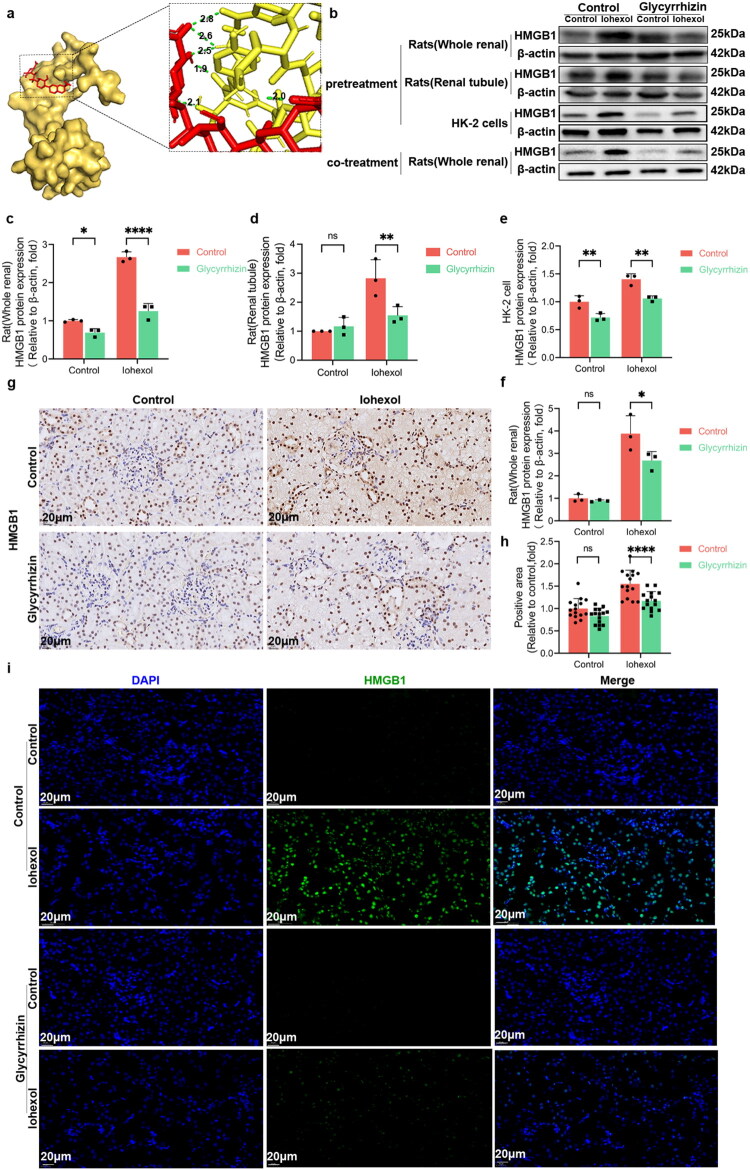
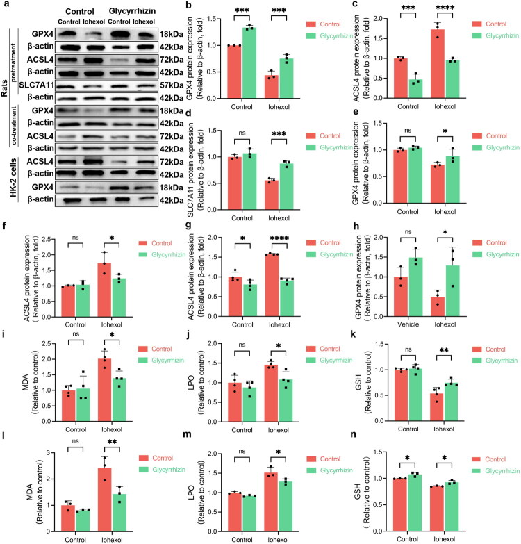
Contrast-induced acute kidney injury (CI-AKI) is the third leading cause of AKI, but there are no effective preventive or therapeutic measures in clinical practice. Glycyrrhizin, a bioactive compound isolated from the Glycyrrhiza glabra L., exhibits anti-inflammatory effects; however, the effects and mechanisms of glycyrrhizin on CI-AKI remain unknown. In present study, the effects of glycyrrhizin on renal dysfunction and tissue damage were evaluated in CI-AKI rats and mice. And the mechanisms were further investigated in iohexol treated renal tubular epithelial cells. Molecular docking and network pharmacology were used to discover the binding targets of glycyrrhizin and identify potential pathogenic pathway. Gene knockout mice and gene silencing cells were used to detect whether glycyrrhizin alleviated CI-AKI through target proteins mediated pathway. Results showed that both pretreatment and co-treatment with glycyrrhizin could alleviate iohexol-induced renal dysfunction and pathological damage in vivo. Similarly, glycyrrhizin could improve iohexol-induced decrease in cell viability of both HK-2 cells and primary mice renal tubular epithelial cells. Mechanistically, glycyrrhizin could directly bind to the active site of HMGB1, then blocking iohexol-induced ferroptosis of renal tubular epithelial cells. HMGB1 silencing was able to inhibit overactivation of AMPK/Beclin-1 axis during CI-AKI, and iohexol-downregulated protein expressions of GPX4 and SLC7A11 were reversed in kidneys of AMPK knockout mice. Comparable results were obtained in vitro with AICAR treatment. Our study is the first to demonstrate that glycyrrhizin exerts both protective and therapeutic effect on CI-AKI by inhibiting tubular epithelial cell ferroptosis via HMGB1/AMPK/Beclin-1 axis, providing a potential choice for treating CI-AKI.

Keywords: Contrast-induced AKI; HMGB1; ferroptosis; glycyrrhizin; renal tubular epithelial cells.


Conflict of interest statement

No potential conflict of interest was reported by the authors.

Figures

Figure 1.
Figure 1.
Iohexol significantly induces renal ferroptosis in vivo. (a) Protocol of the experiments to construct CI-AKI rats and mice models. (b) Representative images of H&E-stained kidneys of CI-AKI rats and mice with or without iohexol treatment. The orange, blue, and green arrows indicate the glomerular, tubular, and interstitial damage, respectively. (c-e) SCr, BUN, and renal tubular injury scores of CI-AKI rats. (f-h) SCr, BUN, and renal tubular injury scores of CI-AKI mice. (i, j) The protein expressions of GPX4, SLC7A11 and ACSL4 in the kidneys of CI-AKI rats. (i, k) The protein expression of GPX4 in the kidneys of CI-AKI mice. (l-n) MDA, LPO, and GSH levels in the kidneys of CI-AKI rats. *p < 0.05, **p < 0.01, ***p < 0.001, ****p < 0.0001. Data are shown as mean ± SD; n = 3 ∼ 4 independent experimental samples per group.
Figure 2.
Figure 2.
Iohexol significantly induces ferroptosis in HK-2 cells, which can be mitigated by Fer-1. (a) Representative images of transmission electron microscopy of HK-2 cells with or without iohexol treatment. Red, yellow, and green arrows indicate mitochondria, nucleus, and lysosome, respectively. (b, c) The protein expressions of GPX4, ACSL4, and Tfr1 in HK-2 cells with or without iohexol treatment. (d) Intracellular levels of MDA, LPO, and GSH in HK-2 cells with or without iohexol treatment. (e) Representative images of intracellular Fe2+ in HK-2 cells treated with iohexol. Orange fluorescence indicates Fe2+. Scale, 100 μm. (f-h) The protein expressions of GPX4 and Tfr1 in HK-2 cells cultured with or without Fer-1 or iohexol. (i, j) Intracellular levels of MDA and GSH in HK-2 cells cultured with or without Fer-1 or iohexol. **p < 0.01, ***p < 0.001, ****p < 0.0001. Data are shown as mean ± SD; n = 3 ∼ 4 independent experimental samples per group.
Figure 3.
Figure 3.
HMGB1 was up-regulated in the kidneys of CI-AKI rats, and HMGB1 silencing alleviated iohexol-induced ferroptosis of HK-2 cells. (a, b) The protein expressions of HMGB1 in rats, mice, or HK-2 cells with or without iohexol treatment. (c) Representative images of immunohistochemistry staining of HMGB1 in the kidneys of CI-AKI rats. (d) Representative images of IF of HMGB1 in the kidneys of CI-AKI rats. (e-h) The protein expressions of HMGB1, GPX4, and ACSL4 in HK-2 cells transfected with or without HMGB1 siRNA or iohexol. (i) Intracellular level of GSH in HK-2 cell models after HMGB1 silencing. *p < 0.05, **p < 0.01, ***p < 0.001, ***p < 0.0001. Data are shown as mean ± SD; n = 3 ∼ 4 independent experimental samples per group.
Figure 4.
Figure 4.
Both pretreatment and co-treatment with glycyrrhizin significantly protect against the kidney injury induced by iohexol. (a) Schematic diagram of glycyrrhizin administration to CI-AKI rats. (b) Representative images of H&E-stained kidneys of CI-AKI rats with or without glycyrrhizin pre-intervention. The orange, blue, and green arrows indicate the glomerular, tubular, and interstitial damage, respectively. (c-e) SCr, BUN, and renal tubular injury scores of CI-AKI rats pretreated with or without glycyrrhizin. (f) Representative images of H&E-stained kidneys of CI-AKI rats with or without glycyrrhizin co-treatment. The orange, blue, and green arrows indicate the glomerular, tubular, and interstitial damage, respectively. (g-i) SCr, BUN, and renal tubular injury scores of CI-AKI rats co-treated with or without glycyrrhizin. (j-k) The cell viability was evaluated with CCK8 in HK-2 cells (j) and primary mice renal tubular epithelial cells (k) co-cultured with or without iohexol and different concentrations of glycyrrhizin for 48h, respectively. *p < 0.05, **p < 0.01, ***p < 0.001, ****p < 0.0001. Data are shown as mean ± SD; n = 3 ∼ 5 independent experimental samples per group.
Figure 5.
Figure 5.
Glycyrrhizin significantly inhibits HMGB1 expression. (a) Molecular docking between glycyrrhizin and HMGB1. (b-e) The protein expressions of HMGB1 in the kidneys or renal tubules of CI-AKI rats, or HK-2 cells with or without glycyrrhizin pretreatment. (b, f) The protein expression of HMGB1 in the kidneys of CI-AKI rats with or without glycyrrhizin co-treatment. (g) Representative IHC images of HMGB1 in the kidneys of CI-AKI rats with or without glycyrrhizin pretreatment. (h) Fifteen fields of view were randomly selected for each slide in same tissue by used Image J software. The positive area in each field was determined using ImageJ software (i) Representative IF images of HMGB1 in the kidneys of CI-AKI rats with or without glycyrrhizin pretreatment. Control-Control image and Control-Iohexol image were derived from the same tissue but different field in same group which described in Figure 3-d.*p < 0.05, **p < 0.01, ****p < 0.0001. Data are shown as mean ± SD; n = 3∼15 independent experimental samples per group.
Figure 6.
Figure 6.
The protective effect of glycyrrhizin on CI-AKI may be partly through inhibiting ferroptosis. (a–d) The protein expressions of GPX4, ACSL4, and SLC7A11 in the kidneys of CI-AKI rats with or without glycyrrhizin pre-intervention. (a, e, f) The protein expressions of ACSL4 and GPX4 in the kidneys of CI-AKI rats with or without glycyrrhizin co-intervention. (a, g, h) The protein expressions of GPX4 and ACSL4 in CI-AKI HK-2 cells with or without glycyrrhizin pre-intervention. (l–k) Levels of MDA, LPO and GSH in the kidney of CI-AKI rats pretreated with glycyrrhizin. (l–n) Levels of MDA, LPO and GSH in CI-AKI HK-2 cells with or without glycyrrhizin pre-intervention. *p < 0.05, **p < 0.01, ***p < 0.001, ****p < 0.0001. Data are shown as mean ± SD; n = 3∼4 independent experimental samples per group.
Figure 7.
Figure 7.
Activation of AMPK/Beclin-1 pathway is involved in the HMGB1 mediated ferroptosis in iohexol treated HK-2 cells. (a) Protein-protein docking between HMGB1(8i9m) and AMPK(4cff). (b–f) The protein expressions of p-AMPK (Thr172), AMPK, p-Beclin-1(Ser93) and Beclin-1 in HK-2 cells with or without iohexol treatment. (g–k) The protein expressions of p-AMPK (Thr172), AMPK, p-Beclin-1(Ser93) and Beclin-1 in HK-2 cells transfected with or without HMGB1 siRNA or iohexol. *p < 0.05, **p < 0.01, ***p < 0.001, ****p < 0.0001. Data are shown as mean ± SD; n = 3 independent experimental samples per group.
Figure 8.
Figure 8.
The effects of AMPK knockout on iohexol-induced renal dysfunction, tissue damage, and protein expressions of GPX4, SLC7A11, and ACSL4 in mice. (a) Breeding and genotyping strategy for generating AMPKα2 knockout mice. (b) Representative images of H&E-stained kidneys of AMPKα2 knockout mice with or without iohexol treatment. The orange, blue, and green arrows indicate the glomerular, tubular, and interstitial damage, respectively. (c–e) SCr, BUN, and renal tubular injury scores of AMPKα2 knockout mice with or without iohexol treatment. (f–i) Protein expressions of GPX4, SLC7A11, and ACSL4 in kidneys of WT or AMPKα2 knockout mice with or without iohexol treatment. *p < 0.05, **p < 0.01, ***p < 0.001, ****p < 0.0001. Data are shown as mean ± SD; n = 3 ∼ 4 independent experimental samples per group.

Similar articles

References

    1. Mccullough PA, Choi JP, Feghali GA, et al. Contrast-induced acute kidney injury. J Am Coll Cardiol. 2016;68(13):1465–1473. doi: 10.1016/j.jacc.2016.05.099. - DOI - PubMed
    1. Yang L, Xing G, Wang L, et al. Acute kidney injury in China: a cross-sectional survey. Lancet. 2015;386(10002):1465–1471. - PubMed
    1. Mcdonald JS, Mcdonald RJ, Carter RE, et al. Risk of intravenous contrast material-mediated acute kidney injury: a propensity score-matched study stratified by baseline-estimated glomerular filtration rate. Radiology. 2014;271(1):65–73. doi: 10.1148/radiol.13130775. - DOI - PubMed
    1. Khandy AH, Shiekh R, Nabi T, et al. Incidence, determinants, and outcome of contrast-induced acute kidney injury following percutaneous coronary intervention at a tertiary care hospital. Saudi J Kidney Dis Transpl. 2023;34(3):214–223. doi: 10.4103/1319-2442.393994. - DOI - PubMed
    1. Feng Y, Jun M, Wang AY, et al. Predicting contrast-associated acute kidney injury. JAMA Netw Open. 2025;8(3):e250107. doi: 10.1001/jamanetworkopen.2025.0107. - DOI - PMC - PubMed

MeSH terms

LinkOut - more resources