Review
. 2025 Sep 1;10(1):282.
doi: 10.1038/s41392-025-02346-0.

Arterial stiffness and vascular aging: mechanisms, prevention, and therapy

Affiliations
Review

Arterial stiffness and vascular aging: mechanisms, prevention, and therapy

Maximilian Jonathan Herzog et al. Signal Transduct Target Ther. .

Abstract

Cardiovascular diseases are the leading cause of morbidity and mortality worldwide. The central underlying mechanisms of cardiovascular diseases are vascular aging and associated arterial stiffness. Arterial stiffness is characterized by structural (e.g., tunica media calcification, alterations in vascular smooth muscle cells, and fibrosis) and functional (e.g., loss of Windkessel function, elevated pulse pressure, and development of isolated systolic hypertension) vascular changes that cause microvascular dysfunction and end-organ damage (e.g., heart failure, vascular dementia, hypertensive retinopathy, and chronic kidney disease). Current research indicates that arterial stiffness is an independent risk factor for cardiovascular diseases and represents a potential target for personalized prevention and therapeutic approaches. In this review, we summarize the pathophysiological mechanisms of vascular aging and arterial stiffness, outline the resulting end-organ damage, present different methods for the measurement of arterial stiffness, highlight the potential role of prevention and therapy, and provide future perspectives for arterial stiffness research. The purpose of this review is to provide a state-of-the-art interdisciplinary and translational approach to arterial stiffness, highlighting unique pathophysiological mechanisms (e.g., perivascular adipose tissue, extracellular vesicles), clinical relevance, and future directions.


Conflict of interest statement

Competing interests: The authors declare no competing interests.

Figures

Fig. 1
Fig. 1
Schematic illustration of the structural and functional differences in atherosclerosis (left) and arterial stiffness (right). VSMC vascular smooth muscle cells, EV extracellular vesicles, ECM extracellular matrix. Created with BioRender.com
Fig. 2
Fig. 2
a Anatomical structure of the vascular wall from a cross-sectional view. The vascular wall is distinguished into the tunica intima, tunica media, and tunica adventitia/externa. Additionally, arteries are surrounded by PVAT. b Structural and functional differences in muscular arteries, elastic arteries, and arterioles. c Age affects arterial structure: we highlight the main alterations in the vascular wall leading to vascular aging and arterial stiffness. d Overview of the underlying molecular and cellular mechanisms of arterial stiffness in the different layers of the arterial system. Created with BioRender.com
Fig. 3
Fig. 3
Low-grade systematic induction of endothelial dysfunction and resulting arterial stiffness. Chronic low-grade inflammation causes a plethora of endothelial molecular mechanisms, such as increased reactive oxygen species (ROS) and decreased activation of endothelial nitric oxide synthase (eNOS), resulting in reduced nitric oxide (NO) bioavailability, impaired barrier function, and transmigration of immune cells. The activation of the renin‒angiotensin‒aldosterone system (RAAS) increases through mineralocorticoid receptor (MR) NADPH oxidase, resulting in the generation of reactive oxygen species (ROS). Furthermore, mineralocorticoid receptor (MR)-induced activation of serum/glucocorticoid regulated kinase 1 (SGK1) causes increased Na+ influx through the endothelial Na+ channel (EnNaC). Elevated intracellular Na+ is associated with F-actin polymerization and endothelial stiffness. Decreased NO bioavailability causes the expression of adhesion molecules (e.g., VCAM and ICAM-1), resulting in the recruitment of monocytes and Th1 cells. Furthermore, decreased NO bioavailability causes transglutaminase 2 (TG2) activation, macrophage activation, and oxidative stress. TG2 is associated with ECM degradation and resulting ECM stiffness. Additionally, severe oxidative stress causes mitochondrial dysfunction and DNA alterations. Dysfunctional perivascular adipose tissue (PVAT) significantly contributes to the inflammatory state. While physiologically providing anti-inflammatory and vasodilatory signaling, age-associated PVAT releases several deteriorating molecules (e.g., leptin, chemerin, visfatin, resistin, and MCP-1). Created with BioRender.com
Fig. 4
Fig. 4
Interaction of endothelial dysfunction and hypercoagulability. Senescent endothelial cells and vascular smooth muscle cells are associated with increased synthesis of procovWFagulant (e.g., von Willebrand factor, fibrinogen) and anticoagulant (tissue factor pathway inhibitor) factors. In addition, hemodynamic forces, proinflammatory mediators and the autonomic state influence coagulation, resulting in increased endothelial cell permeability and perivascular inflammation. Created with BioRender.com
Fig. 5
Fig. 5
a Pulse wave analysis in a compliant versus a stiff arterial system. A compliant aorta is characterized by sufficient Windkessel function and dampened pulsatility. In contrast, a stiff aorta loses its Windkessel function, resulting in high pulsatility toward the periphery and a wave back unfavorably augmenting the central systolic pressure (augmentation pressure). b Pulse wave analysis of the carotid artery. The pressure decay during diastole can be approximated by the product of peripheral resistance (R) and arterial compliance c. c The first graph (I) depicts the proteins carrying the load at a certain pressure condition. Under hypertensive circumstances, collagen bears the load, so the vessel is not able to expand significantly, whereas under low pressure, elastin enables sufficient stretching qualities. The second graph (II) depicts distensibility properties in addition to the pulse wave velocity (PWV). Both curves function in a nonlinear and nonproportional manner, suggesting that hypertension leads to increased arterial stiffness. Created with BioRender.com
Fig. 6
Fig. 6
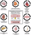
Effects of arterial stiffness on peripheral end organs and clinical manifestations. Arterial stiffening tends to create a vicious cycle in each organ. EPVS enlarged perivascular space, CSVD cerebral small vessel disease, HFpEF heart failure with preserved ejection fraction, MASLD metabolic dysfunction-associated steatotic liver disease, PWV, pulse wave velocity, PAD peripheral artery disease. Created with BioRender.com
Fig. 7
Fig. 7
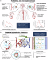
a Microscopic and macroscopic consequences of arterial stiffness and altered vessel pulsation on the brain parenchyma. The increased pressure peaks cause a remodeling process in the arterial wall structure; thus, pulsation is not transmitted into the peripheral microvasculature. This leads to a decrease in CSF influx, the deposition of amyloid proteins and the emergence of enlarged perivascular spaces. In addition, the remodeling process includes decreases in cerebral perfusion and small vessel disease, which can be visualized as microbleeds, white matter hyperintensities, and lacunar infarctions., b Impaired glymphatic clearance is presented on a microscopic scale. The altered pulsation caused by vessel remodeling and arterial stiffness leads to decreased CSF influx into the brain parenchyma and subsequent waste deposition, such as that of amyloid proteins. Stored amyloids cause inflammation and reactive astrogliosis, which promote neurodegenerative diseases. Additionally, we depicted the ongoing process in young individuals versus Alzheimer’s disease patients with enlarged perivascular spaces. CSF cerebrospinal fluid, ISF interstitial fluid, EPVS enlarged perivascular space, pp pulse pressure. Created with BioRender.com
Fig. 8
Fig. 8
Arterial stiffness framework. Future translational arterial stiffness research should (i) enhance clinical assessment, (ii) identify specific phenotypes and endotypes, (iii) decipher the underlying mechanisms and “new” risk factors (e.g., chronic psychosocial stress, air pollution), and (iv) develop tailored prevention and therapy approaches. Created with BioRender.com

References

    1. Gaziano, T., Reddy, K. S., Paccaud, F., Horton, S. & Chaturvedi, V. Cardiovascular Disease. in Disease Control Priorities in Developing Countries. 2nd edition (The International Bank for Reconstruction and Development / The World Bank, 2006). - PubMed
    1. Cardiovascular diseases (CVDs). https://www.who.int/news-room/fact-sheets/detail/cardiovascular-diseases... (2021).
    1. Fishbein, M. C. & Fishbein, G. A. Arteriosclerosis: facts and fancy. Cardiovasc. Pathol.24, 335–342 (2015). - PubMed
    1. Climie, R. et al. Vascular ageing: moving from bench towards bedside. Eur. J. Prev. Cardiol. 30, 1101−1117 (2023). - PMC - PubMed
    1. Fishbein, G. A. & Fishbein, M. C. Arteriosclerosis: rethinking the current classification. Arch. Pathol. Lab. Med.133, 1309–1316 (2009). - PubMed

MeSH terms

LinkOut - more resources