. 2025 Aug 31;26(1):265.
doi: 10.1186/s12931-025-03340-4.

The LPAR1 antagonist, PIPE-791 produces antifibrotic effects in models of lung fibrosis

Affiliations

The LPAR1 antagonist, PIPE-791 produces antifibrotic effects in models of lung fibrosis

Michael Poon et al. Respir Res. .

Abstract

Background: Idiopathic pulmonary fibrosis (IPF) is a chronic progressive form of interstitial lung disease (ILD) characterized by significant extracellular matrix deposition, alveolar damage, and tissue remodeling. Antagonists against the G-protein coupled receptor, lysophosphatidic acid receptor 1 (LPAR1) have shown efficacy in lung fibrosis preclinically and clinically. Here, we profile PIPE-791, a small molecule, orally bioavailable LPAR1 receptor antagonist, and show its effectiveness in several lung fibrosis-related contexts.

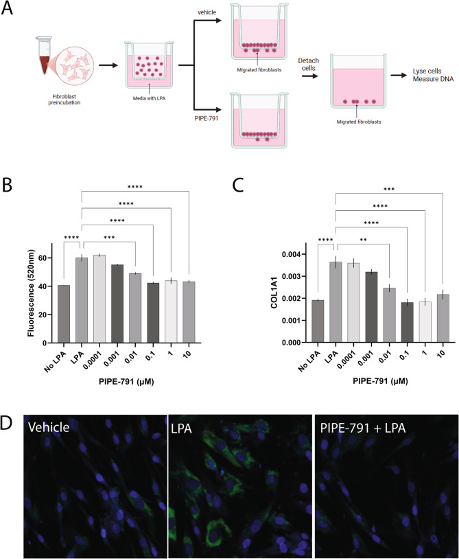
Methods: In vitro, we used human lung fibroblasts and precision cut lung slices (PCLS) derived from donors with pulmonary fibrosis to test PIPE-791 efficacy in reducing markers of fibrosis. In vivo, we used bleomycin-induced lung fibrosis models to demonstrate PIPE-791 efficacy.

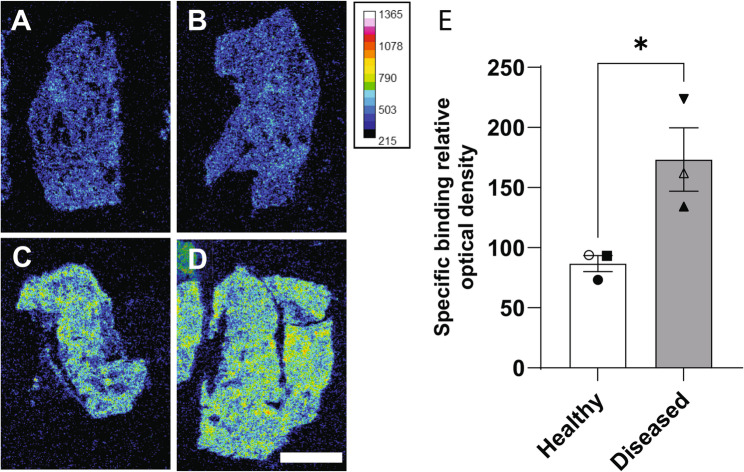
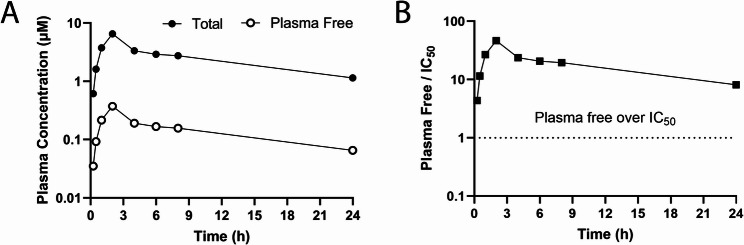
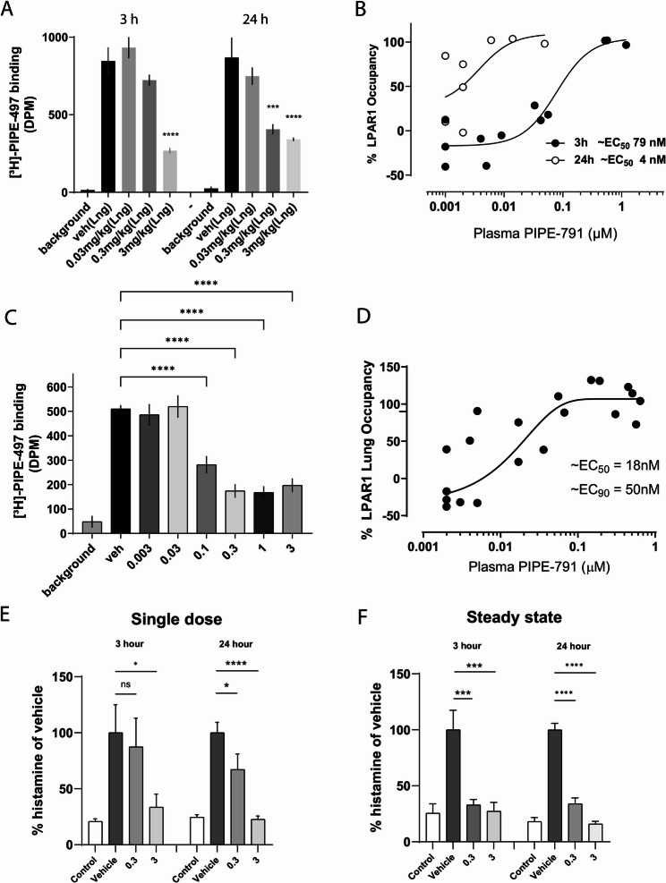
Results: In vitro PIPE-791 reduced LPA-induced collagen expression (IC50 1.1 nM) in human lung fibroblasts. We also show that LPAR1 is elevated in IPF lung tissue and that PIPE-791 significantly reduced several markers of lung fibrosis in PCLS as measured by gene expression and secreted biomarkers. Using in vivo receptor occupancy, we found that PIPE-791 has long association kinetics resulting in a 20-fold increase in potency when dosed 3 versus 24 h prior to radioligand administration. At 3 mg/kg, PIPE-791 was effective in significantly reducing markers of fibrosis and collagen expression in mouse bleomycin models.

Conclusions: We show that PIPE-791 effectively reduces fibrosis and fibrotic markers in vitro and in vivo and that it has slow association and dissociation kinetics. Taken together, our data support clinical testing of PIPE-791 in the context of fibrotic conditions such as IPF.


Conflict of interest statement

Declarations. Ethics approval and consent to participate: Precision cut lung slices were obtained from Anabios (San Diego, CA) and collected in accordance with their ethics policy ( https://anabios.com/ethics-statement/ ) or approved by the Hanover medical school ethics committee (Hanover, Germany) and approved by Contineum Therapeutics. All animal procedures were approved by the Contineum Therapeutics Institutional Animal Care and Use Committee. Consent for publication: Not applicable. Competing interests: MP, KL, AB, KS, DB, GE, GJ, CB, JR, TS, LV, YX, AC, and DL are current or former employees of Contineum Therapeutics. All authors hold financial shares of Contineum Therapeutics. Contineum Therapeutics owns patent rights to PIPE-791.

Figures

Fig. 1
Fig. 1
PIPE-791 inhibits collagen expression and chemotaxis in primary adult human lung fibroblasts. A. Diagram outlining the chemotaxis assay: human lung fibroblasts are incubated with PIPE-791 and plated onto a Transwell culture insert with LPA in bottom chamber as a chemoattractant. Fibroblasts migrate through the pores and along the bottom surface. Cells on the bottom surface are detached, lysed and the lysate analyzed for DNA content with DAPI. B. Concentration dependent inhibition of LPA-induced chemotaxis as measured by DAPI fluorescence (A520 nm). PIPE-791 inhibits chemotaxis at an IC50 of 1.5 nM (n=3, ** P < 0.01, *** P < 0.001, **** P < 0.0001, one-way ANOVA, error bars SEM). C. Concentration dependent inhibition of LPA induced COL1A1 expression in fibroblasts. PIPE-791 inhibits COL1A1 induction with an IC50 of 1.1 nM (n=3, ** P < 0.01, *** P < 0.001, *** P < 0.0001, one-way ANOVA, Newman-Keuls, error bars SEM). D. Representative images of primary human lung fibroblasts treated with vehicle, LPA, or PIPE-791 and LPA and stained for COL1A1 (green) and Hoechst (blue). Each micrograph is 100 µm width
Fig. 2
Fig. 2
PIPE-791 inhibits TGFβ1 or LPA-induced myofibroblast transformation. Human adult fibroblasts were treated and co-stained for αSMA and COL1A1. A. Representative images of fibroblasts stained with αSMA (white) after treating with vehicle, TGFβ1, or PIPE-791/TGFβ (top row) or LPA, PIPE-791/LPA. A1. PIPE-791 (1 μM) reduced αSMA at all concentrations of TGFβ1 or (A2) LPA tested. B. Representative images of primary human lung fibroblasts stained for COL1A1 (green) after treating with vehicle, TGFβ1, or PIPE-791/ TGFβ (top row), or LPA, PIPE-791/LPA. B1. PIPE-791 (1 μM) reduced COL1A1 expression at all concentrations of TGFβ1 or (B2) LPA tested (n  >  5, * P < 0.05, ** P < 0.01, *** P < 0.001, **** P < 0.0001, t-test, error bars SEM)
Fig. 3
Fig. 3
LPAR1 is more highly expressed in diseased lung tissue. A-D. Representative images illustrating the total binding of [3H]-PIPE-497 (20 nM) on human lung sections from healthy (A, B) and diseased lungs (C, D). Intensity scale of the relative optical density is shown. Scale bar = 0.5 mm. E. Bar graph with individual values showing increased in LPAR1 specific binding signal in diseased compared to healthy donors. (n = 3 for healthy and diseased tissue, * P < 0.05, t-test, error bars SD). Symbols correspond to donors in Additional File 1 as follows: ⬤ Donor A, ◯ Donor B, ⬛ Donor C, ▲ Donor D, △ Donor E, ▼Donor F
Fig. 4
Fig. 4
PIPE-791 reduces expression of fibrotic markers in human PCLS. A-F. Six day incubation of PIPE-791 with PCLS from donors with pulmonary fibrosis decreases COL1A1 expression (A), COL3A1 (B), SERPINE1 (C), and TIMP1 (D) (n = 6 donors, * P < 0.05, ** P < 0.01, t-test, error bars SD). E. PIPE-791 does not significantly change LPAR1 expression (n = 6, n.s. not significant, t-test). F. PIPE-791 does not impact slice health compared to vehicle treated PCLS as measured by LDH in the media (n = 6, n.s. not significant, t-test). Symbols correspond to donors in Additional File 1 as follows: Donor G; ■ Donor H; ▲ Donor I; ◆ Donor J; ⬣ Donor K; ▼ Donor L. G-I. Forty-eight hour incubation of nintedanib or 3 concentrations of PIPE-791 (0.03, 0.3, or 3 µM) with fresh PCLS from donors with pulmonary fibrosis results in significant inhibition of secreted C3M (G) and Pro-C6 (H). Pro-C3 (I) did not show a significant decrease with nintedanib or PIPE-791 (n = 12, two donors, two lung regions in triplicate, * P < 0.05, ** P < 0.01, *** P < 0.001, **** P < 0.0001, one-way ANOVA, Dunnett’s post-hoc, error bars are SD). Donor information can be found in Additional File 1
Fig. 5
Fig. 5
PIPE-791 has extended plasma exposure in rat. A Rats were orally dosed with 10 mg/kg PIPE-791 and plasma concentrations taken at 0.25, 0.5, 1, 2, 4-, 6-, 8-, and 24-hours post dose. Total PIPE-791 concentration (solid circles) and free concentrations (after correcting for a rat plasma fraction unbound of 0.057, open circles) shown, n = 2. B Graph depicting PIPE-791 plasma free (unbound) concentration as a function of time over the in vitro calcium mobilization IC50. At 10 mg/kg, the free PIPE-791 concentration in plasma is still well above its LPAR1 IC50 at the last timepoint measured (24 h).
Fig. 6
Fig. 6
PIPE-791 has long receptor dissociation kinetics and inhibits mast cell histamine release in vivo. A Mice were orally dosed with PIPE-791 followed by intravenous injection of the LPAR1 selective radiotracer [3H]-PIPE-497 at 3 or 24 h post dose and lung collection. Significant radiotracer displacement (LPAR1 receptor occupancy) was observed at 3 mg/kg at 3 h, whereas significant occupancy was observed at both 0.3 and 3 mg/kg at 24 h (n > 4, ANOVA with Newman-Keuls, *** P < 0.001, **** P < 0.0001, error bars SEM). B Values plotted as free plasma concentration versus percent occupancy highlighting increase in EC50 from 79 nM at 3 h to 4 nM at 24 h, 3 h (solid circles) and 24 h (open circles, error bars SEM). C Mice were orally dosed PIPE-791 for 4 days, once a day to simulate steady state conditions. Lung occupancy was measured by i.v. injection of [3H]-PIPE-497. Lung receptor occupancy gave an ED50 of 0.1 mg/kg (n > 5, except background n = 4, **** P < 0.0001, One way ANOVA, with Tukey’s, error bars SEM). D Based on free plasma concentrations, the calculated EC50 and EC90 were 18 nM and 50 nM, respectively. E Mice were orally dosed with PIPE-791 at 0.3 or 3 mg/kg then challenged with intravenous LPA 3–24 h later. Blood histamine levels were analyzed. Significant reduction in histamine was observed at 3 mg/kg at 3 h and at both 0.3 and 3 mg/kg at 24 h (n > 4 for all groups except vehicle at 3 h n = 2; ANOVA, uncorrected Fisher’s LSD, * P < 0.05, **** P < 0.0001, error bars SEM). F Mice were dosed with PIPE-791 for 4 days to achieve steady-state followed by LPA challenge and blood histamine collection at 3 and 24 h post last dose. Significant reduction at both 0.3 and 3 mg/kg at both 3 and 24 h (n = 6, ANOVA with Newman-Keuls, *** P < 0.001, **** P < 0.0001, error bars SEM)
Fig. 7
Fig. 7
PIPE-791 reduces fibrotic markers in an intratracheal bleomycin model. A Mice treated with PIPE-791 show dose dependent body weight improvement compared to vehicle/bleomycin (Veh/BLEO) treated mice (n = 8, deaths excluded, one way ANOVA at day 14, ** P < 0.01, *** P < 0.001, error bars are SEM). B Survival plot of study over time. No deaths were observed in the PIPE-791 treated groups. C BAL fluid was collected at study termination and analyzed for fibrotic markers. PIPE-791 significantly inhibited several markers in the presence of bleomycin in a dose dependent fashion including procollagen, (D) TGFβ1, and (E) TIMP1 (n = 8, deaths excluded, ANOVA with Newman-Keuls, * P < 0.05, ** P < 0.01, *** P < 0.001, **** P < 0.0001, error bars SEM). F PIPE-791 reduced collagen amounts in lung tissue (n = 8, excluding deaths, one-way ANOVA with Newman Keuls, * P < 0.05, **** P < 0.0001, error bars SEM)
Fig. 8
Fig. 8
Therapeutic dosing of PIPE-791 reduces fibrotic markers in a systemic bleomycin model. A Representative trichrome stained images of vehicle/vehicle, bleomycin/vehicle, bleomycin/BMS-986020, and bleomycin/PIPE-791 treated mice (blue, collagen). BMS-986020 was used as a positive control LPAR1 antagonist. Right column is magnified image of region in white box. B PIPE-791 significantly reduces lung tissue collagen fraction as assessed histologically with Maisson’s trichrome staining (blue positive area/total tissue area x 100%; veh n = 8, Bleo/vehicle n = 11, Bleo/BMS-986020 n = 12, Bleo/PIPE-791 n = 12, * P < 0.05, ** P < 0.01, n.s. non-significant, one-way ANOVA with Tukey’s, error bars SEM). C PIPE-791 reduces bleomycin induced lung hydroxyproline levels (veh n = 8, Bleo/vehicle n = 11, Bleo/BMS-986020 n = 12, Bleo/PIPE-791 n = 12, * P < 0.05, one-way ANOVA with Tukey’s, error bars SEM).
Fig. 9
Fig. 9
PIPE-791 regulates macrophage activation and release of IL-1β. A Primary human alveolar macrophages were treated with PIPE-791 then stimulated ex vivo with LPS. PIPE-791 significantly reduced IL-1 secretion into the culture media (n > 5, * P < 0.05, one-way ANOVA with Newman-Keuls, error bars SEM). B Alveolar macrophages were collected from mice orally dosed in vivo with 3 mg/kg PIPE-791 followed by ex vivo stimulation with LPS. Treatment with PIPE-791 reduced secreted IL-1β levels (n > 9, one-way ANOVA with Newman-Keuls, * P < 0.05, error bars SEM). C Bleomycin was administered intratracheally, then mice orally dosed with 3 mg/kg PIPE-791. BAL fluid was collected, separated into pellet and supernatant fractions, and analyzed. Cells were stained with the macrophage marker CD68, showing an increase in size (an indicator of activation) in response to bleomycin. Activation was quantified as the mean area of CD68 staining per cell. Bleomycin increased cell size which was reversed in mice dosed orally with PIPE-791 (n = 5, one-way ANOVA with Tukey’s, * P < 0.05, ** P < 0.01, **** P < 0.0001, error bars SEM). D Representative images of cells collected from BAL fluid in (C) stained with the macrophage marker CD68 (green, left column) showing an increase in size (an indicator of activation) in response to LPS stimulation. Thresholded images (right column). E Analysis of IL-1β in the BAL fluid showed a significant reduction in bleomycin induced IL-1β in mice dosed with PIPE-791 (n = 4, one-way ANOVA with Tukey’s, ** P < 0.01, *** P < 0.001, error bars SEM)

References

    1. Raghu G, Remy-Jardin M, Richeldi L, Thomson CC, Inoue Y, Johkoh T, Kreuter M, Lynch DA, Maher TM, Martinez FJ, et al. Idiopathic pulmonary fibrosis (an Update) and progressive pulmonary fibrosis in adults: an official ATS/ERS/JRS/ALAT clinical practice guideline. Am J Respir Crit Care Med. 2022;205:e18–47. - PMC - PubMed
    1. Selman M, Pardo A. The leading role of epithelial cells in the pathogenesis of idiopathic pulmonary fibrosis. Cell Signal. 2020;66:109482. - PubMed
    1. Glass DS, Grossfeld D, Renna HA, Agarwala P, Spiegler P, DeLeon J, Reiss AB. Idiopathic pulmonary fibrosis: current and future treatment. Clin Respir J. 2022;16:84–96. - PMC - PubMed
    1. Swaney JS, Chapman C, Correa LD, Stebbins KJ, Bundey RA, Prodanovich PC, Fagan P, Baccei CS, Santini AM, Hutchinson JH, et al. A novel, orally active LPA(1) receptor antagonist inhibits lung fibrosis in the mouse bleomycin model. Br J Pharmacol. 2010;160:1699–713. - PMC - PubMed
    1. Tager AM, LaCamera P, Shea BS, Campanella GS, Selman M, Zhao Z, Polosukhin V, Wain J, Karimi-Shah BA, Kim ND, et al. The lysophosphatidic acid receptor LPA1 links pulmonary fibrosis to lung injury by mediating fibroblast recruitment and vascular leak. Nat Med. 2008;14:45–54. - PubMed

MeSH terms

LinkOut - more resources