

HIF-1α: a bridge connecting sepsis and acute respiratory distress syndrome
- PMID: 40887582
- PMCID: PMC12399009
- DOI: 10.1186/s40001-025-03107-z
HIF-1α: a bridge connecting sepsis and acute respiratory distress syndrome
Abstract
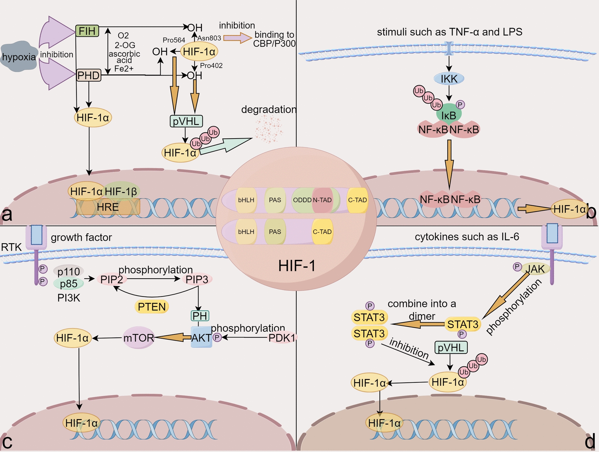
Sepsis is a life-threatening condition marked by an abnormal host response to infection that can result in organ dysfunction, making it recognized as one of the primary causes of acute respiratory distress syndrome (ARDS). The pathophysiology of sepsis involves a cascade of events, including heightened pulmonary capillary permeability, dysfunction of alveolar epithelial cells, and the infiltration of inflammatory cells, such as neutrophils, macrophages, monocytes, and lymphocytes. The presence of these inflammatory cells triggers capillary leakages, alveolar epithelial damage, and the accumulation of fluid within the alveolar spaces, leading to compromised gas exchange, acute respiratory failure, and the progression to ARDS. In this complex scenario, Hypoxia-Inducible Factor-1α (HIF-1α) emerges as a pivotal player in maintaining cellular oxygen homeostasis and responding to hypoxia and inflammatory stimuli. This narrative review delves into the intricate molecular and biological characteristics of HIF-1α, elucidating its regulatory role within the context of sepsis and ARDS. By exploring the therapeutic potential of targeting HIF-1α, this review seeks to offer valuable insights into the underlying mechanisms linking sepsis to ARDS. Ultimately, this exploration of HIF-1α seeks to enhance our comprehension of sepsis pathogenesis, identify novel therapeutic avenues, and lay a strong theoretical groundwork for future clinical interventions.
Keywords: Acute respiratory distress syndrome; Critical care; HIF-1α; Hypoxia; Molecular medicine; Sepsis.
© 2025. The Author(s).
Conflict of interest statement
Declarations. Ethics approval and consent to participate: Not applicable. Consent for publication: Not applicable. Competing interests: The authors declare no competing interests.
Figures


Similar articles
-
Systemic Inflammatory Response Syndrome.2025 Jun 20. In: StatPearls [Internet]. Treasure Island (FL): StatPearls Publishing; 2025 Jan–. 2025 Jun 20. In: StatPearls [Internet]. Treasure Island (FL): StatPearls Publishing; 2025 Jan–. PMID: 31613449 Free Books & Documents.
-
Ferroptosis: a key driver and therapeutic target in the pathogenesis of acute respiratory distress syndrome.Front Immunol. 2025 Jul 22;16:1567980. doi: 10.3389/fimmu.2025.1567980. eCollection 2025. Front Immunol. 2025. PMID: 40766305 Free PMC article. Review.
-
CGRP alleviates lipopolysaccharide-induced ARDS inflammation via the HIF-1α signaling pathway.Clin Sci (Lond). 2025 Apr 9;139(7):373-387. doi: 10.1042/CS20243170. Clin Sci (Lond). 2025. PMID: 40110637 Free PMC article.
-
Rabeprazole Promotes Vascular Repair and Resolution of Sepsis-Induced Inflammatory Lung Injury through HIF-1α.Cells. 2022 Apr 22;11(9):1425. doi: 10.3390/cells11091425. Cells. 2022. PMID: 35563731 Free PMC article.
-
Exploring the role of HIF-1α in pathogenesis of parkinson's disease and its potential as a therapeutic target.Mol Biol Rep. 2025 Sep 2;52(1):858. doi: 10.1007/s11033-025-10944-y. Mol Biol Rep. 2025. PMID: 40892154 Review.
References
Publication types
MeSH terms
Substances
Grants and funding
LinkOut - more resources
Full Text Sources
Medical
